Introduction
Benign neoplasms of the respiratory system are non-cancerous growths that can occur in various parts of the respiratory tract, including the lungs, bronchi, trachea, and nasal passages. While these growths are not typically life-threatening, they can cause symptoms and complications that may require medical intervention.[1] The aim of this guide is to provide a comprehensive overview of the diagnosis and management of benign neoplasms of the respiratory system.
Codes
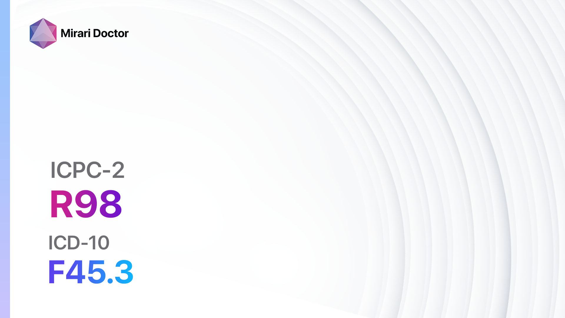
- ICPC-2 Code: R86 Benign neoplasm respiratory
- ICD-10 Code: D14.3 Benign neoplasm of bronchus and lung[2]
Symptoms
- Cough: Persistent cough that may be dry or produce mucus.
- Shortness of breath: Difficulty breathing or a feeling of breathlessness.
- Wheezing: High-pitched whistling sound when breathing.
- Chest pain: Discomfort or pain in the chest.
- Hemoptysis: Coughing up blood.
- Recurrent respiratory infections: Frequent infections of the respiratory tract.[3]
Causes
- Genetic factors: Certain genetic mutations may increase the risk of developing benign neoplasms of the respiratory system.
- Environmental factors: Exposure to certain substances, such as asbestos or tobacco smoke, may increase the risk.
- Chronic inflammation: Conditions that cause chronic inflammation in the respiratory tract, such as chronic bronchitis or sinusitis, may increase the risk.[4]
Diagnostic Steps
Medical History
- Gather information about the patient’s symptoms, including the duration and severity.
- Ask about any risk factors, such as exposure to environmental toxins or a family history of respiratory neoplasms.
- Inquire about any relevant medical conditions, such as chronic respiratory infections or inflammatory diseases.[5]
Physical Examination
- Perform a thorough examination of the respiratory system, including auscultation of the lungs and examination of the nasal passages and throat.
- Look for any signs of respiratory distress, such as increased respiratory rate or use of accessory muscles.
- Check for any palpable masses or lymph node enlargement.[6]
Laboratory Tests
- Complete blood count (CBC): To assess for any abnormalities, such as anemia or infection.
- Pulmonary function tests: To evaluate lung function and assess for any obstructive or restrictive patterns.
- Sputum cytology: Examination of sputum under a microscope to look for abnormal cells.
- Tumor markers: Blood tests to measure specific markers that may be elevated in certain types of respiratory neoplasms.[7]
Diagnostic Imaging
- Chest X-ray: To visualize the lungs and assess for any abnormalities, such as masses or infiltrates.
- Computed tomography (CT) scan: Provides detailed images of the respiratory system and can help determine the location and extent of the neoplasm.
- Magnetic resonance imaging (MRI): May be used to further evaluate the neoplasm and surrounding structures.
- Positron emission tomography (PET) scan: Can help determine if the neoplasm is cancerous or benign by measuring metabolic activity.[8]
Other Tests
- Bronchoscopy: A flexible tube with a camera is inserted into the airways to visualize the neoplasm and collect tissue samples for biopsy.
- Biopsy: Removal of a small sample of tissue for examination under a microscope to determine if the neoplasm is benign or malignant.
- Genetic testing: In some cases, genetic testing may be performed to identify specific mutations associated with respiratory neoplasms.[9]
Follow-up and Patient Education
- Schedule regular follow-up appointments to monitor the neoplasm and assess for any changes or progression.
- Educate the patient about the nature of the neoplasm, its benign nature, and the importance of regular monitoring.
- Provide information about lifestyle modifications and interventions that may help manage symptoms and reduce the risk of complications.[10]
Possible Interventions
Traditional Interventions
Medications:
Top 5 drugs for Benign neoplasm respiratory:
- Corticosteroids (e.g., Prednisone, Dexamethasone):
- Cost: Generic versions can range from $5 to $50 per month.
- Contraindications: Active infections, systemic fungal infections.
- Side effects: Increased appetite, weight gain, mood changes.
- Severe side effects: Increased risk of infections, osteoporosis.
- Drug interactions: Nonsteroidal anti-inflammatory drugs (NSAIDs), certain antibiotics.
- Warning: Long-term use may require gradual tapering to avoid adrenal insufficiency.
- Bronchodilators (e.g., Albuterol, Salmeterol):
- Cost: Generic versions can range from $10 to $50 per month.
- Contraindications: Hypersensitivity to the medication.
- Side effects: Tremor, increased heart rate.
- Severe side effects: Paradoxical bronchospasm, cardiovascular effects.
- Drug interactions: Beta-blockers, certain antidepressants.
- Warning: Overuse may lead to decreased effectiveness.
- Antihistamines (e.g., Loratadine, Cetirizine):
- Cost: Generic versions can range from $5 to $20 per month.
- Contraindications: Hypersensitivity to the medication.
- Side effects: Drowsiness, dry mouth.
- Severe side effects: Rare, but may include allergic reactions.
- Drug interactions: Sedatives, alcohol.
- Warning: May cause drowsiness, avoid activities requiring mental alertness.
- Mucolytics (e.g., Acetylcysteine, Carbocisteine):
- Cost: Generic versions can range from $10 to $30 per month.
- Contraindications: Hypersensitivity to the medication.
- Side effects: Nausea, vomiting.
- Severe side effects: Rare, but may include bronchospasm.
- Drug interactions: None known.
- Warning: Administer with caution in patients with a history of peptic ulcer disease.
- Antibiotics (e.g., Amoxicillin, Azithromycin):
- Cost: Generic versions can range from $5 to $30 per course of treatment.
- Contraindications: Hypersensitivity to the medication.
- Side effects: Nausea, diarrhea.
- Severe side effects: Rare, but may include severe allergic reactions.
- Drug interactions: None known.
- Warning: Use with caution in patients with a history of antibiotic-associated colitis.
Alternative Drugs:
- Leukotriene modifiers (e.g., Montelukast): Can help reduce inflammation in the airways.
- Immunomodulators (e.g., Methotrexate): May be used in certain cases to suppress the immune response.
- Antifungal medications (e.g., Fluconazole): If a fungal infection is suspected or identified.
Surgical Procedures:
- Resection: Surgical removal of the neoplasm may be considered if it is causing significant symptoms or complications.
- Laser therapy: In some cases, laser therapy may be used to remove or shrink the neoplasm.
- Cryotherapy: Freezing the neoplasm with liquid nitrogen to destroy the abnormal tissue.
- Electrocautery: Using an electric current to burn and destroy the neoplasm.
- Photodynamic therapy: A light-sensitive medication is applied to the neoplasm and activated with a laser to destroy the abnormal tissue.
Alternative Interventions
- Acupuncture: May help alleviate symptoms and improve overall well-being. Cost: $60-$120 per session.
- Herbal supplements: Certain herbs, such as turmeric or ginger, may have anti-inflammatory properties. Cost: Varies depending on the specific supplement.
- Breathing exercises: Techniques such as pursed-lip breathing or diaphragmatic breathing may help improve lung function and reduce symptoms. Cost: Free.
- Yoga: Gentle stretching and breathing exercises in yoga may help improve lung capacity and reduce stress. Cost: Varies depending on the location and instructor.
- Meditation: Mindfulness meditation techniques may help reduce stress and improve overall well-being. Cost: Free.
Lifestyle Interventions
- Smoking cessation: Quitting smoking is crucial in managing respiratory neoplasms and reducing the risk of complications. Cost: Varies depending on the method chosen (e.g., nicotine replacement therapy, medications, counseling).
- Regular exercise: Engaging in regular physical activity can help improve lung function and overall fitness. Cost: Varies depending on the chosen activity (e.g., gym membership, home exercise equipment).
- Healthy diet: Consuming a balanced diet rich in fruits, vegetables, and whole grains can support overall health and immune function. Cost: Varies depending on individual food choices.
- Avoidance of environmental toxins: Minimizing exposure to environmental pollutants, such as secondhand smoke or occupational hazards, can help reduce the risk of respiratory neoplasms. Cost: Varies depending on individual circumstances.
- Stress management: Finding effective stress management techniques, such as meditation or counseling, can help improve overall well-being. Cost: Varies depending on the chosen method (e.g., self-guided techniques, professional counseling).
It is important to note that the cost ranges provided are approximate and may vary depending on the location and availability of the interventions. It is recommended to consult with healthcare professionals for personalized recommendations and cost estimates.
Mirari Cold Plasma Alternative Intervention
Understanding Mirari Cold Plasma
- Safe and Non-Invasive Treatment: Mirari Cold Plasma is a safe and non-invasive treatment option for various skin conditions. It does not require incisions, minimizing the risk of scarring, bleeding, or tissue damage.
- Efficient Extraction of Foreign Bodies: Mirari Cold Plasma facilitates the removal of foreign bodies from the skin by degrading and dissociating organic matter, allowing easier access and extraction.
- Pain Reduction and Comfort: Mirari Cold Plasma has a local analgesic effect, providing pain relief during the treatment, making it more comfortable for the patient.
- Reduced Risk of Infection: Mirari Cold Plasma has antimicrobial properties, effectively killing bacteria and reducing the risk of infection.
- Accelerated Healing and Minimal Scarring: Mirari Cold Plasma stimulates wound healing and tissue regeneration, reducing healing time and minimizing the formation of scars.
Mirari Cold Plasma Prescription
Video instructions for using Mirari Cold Plasma Device – R86 Benign neoplasm respiratory (ICD-10:D14.3)
| Mild | Moderate | Severe |
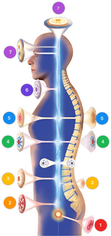
| Mode setting: 1 (Infection) Location: 5 (Lungs) Morning: 15 minutes, Evening: 15 minutes |
Mode setting: 1 (Infection) Location: 5 (Lungs) Morning: 30 minutes, Lunch: 30 minutes, Evening: 30 minutes |
Mode setting: 1 (Infection) Location: 5 (Lungs) Morning: 30 minutes, Lunch: 30 minutes, Evening: 30 minutes |
| Mode setting: 2 (Wound Healing) Location: 5 (Lungs) Morning: 15 minutes, Evening: 15 minutes |
Mode setting: 2 (Wound Healing) Location: 5 (Lungs) Morning: 30 minutes, Lunch: 30 minutes, Evening: 30 minutes |
Mode setting: 2 (Wound Healing) Location: 5 (Lungs) Morning: 30 minutes, Lunch: 30 minutes, Evening: 30 minutes |
| Mode setting: 3 (Antiviral Therapy) Location: 5 (Lungs) Morning: 15 minutes, Evening: 15 minutes |
Mode setting: 3 (Antiviral Therapy) Location: 5 (Lungs) Morning: 30 minutes, Lunch: 30 minutes, Evening: 30 minutes |
Mode setting: 3 (Antiviral Therapy) Location: 5 (Lungs) Morning: 30 minutes, Lunch: 30 minutes, Evening: 30 minutes |
| Mode setting: 7 (Immunotherapy) Location: 4 (Heart, Bile & Pancreas) Morning: 15 minutes, Evening: 15 minutes |
Mode setting: 7 (Immunotherapy) Location: 4 (Heart, Bile & Pancreas) Morning: 30 minutes, Lunch: 30 minutes, Evening: 30 minutes |
Mode setting: 7 (Immunotherapy) Location: 4 (Heart, Bile & Pancreas) Morning: 30 minutes, Lunch: 30 minutes, Evening: 30 minutes |
| Total Morning: 60 minutes approx. $10 USD, Evening: 60 minutes approx. $10 USD |
Total Morning: 120 minutes approx. $20 USD, Lunch: 120 minutes approx. $20 USD, Evening: 120 minutes approx. $20 USD, |
Total Morning: 120 minutes approx. $20 USD, Lunch: 120 minutes approx. $20 USD, Evening: 120 minutes approx. $20 USD, |
| Usual treatment for 7-60 days approx. $140 USD – $1200 USD | Usual treatment for 6-8 weeks approx. $2,520 USD – $3,360 USD |
Usual treatment for 3-6 months approx. $5,400 USD – $10,800 USD
|
 |
|
Use the Mirari Cold Plasma device to treat Benign neoplasm respiratory effectively.
WARNING: MIRARI COLD PLASMA IS DESIGNED FOR THE HUMAN BODY WITHOUT ANY ARTIFICIAL OR THIRD PARTY PRODUCTS. USE OF OTHER PRODUCTS IN COMBINATION WITH MIRARI COLD PLASMA MAY CAUSE UNPREDICTABLE EFFECTS, HARM OR INJURY. PLEASE CONSULT A MEDICAL PROFESSIONAL BEFORE COMBINING ANY OTHER PRODUCTS WITH USE OF MIRARI.
Step 1: Cleanse the Skin
- Start by cleaning the affected area of the skin with a gentle cleanser or mild soap and water. Gently pat the area dry with a clean towel.
Step 2: Prepare the Mirari Cold Plasma device
- Ensure that the Mirari Cold Plasma device is fully charged or has fresh batteries as per the manufacturer’s instructions. Make sure the device is clean and in good working condition.
- Switch on the Mirari device using the power button or by following the specific instructions provided with the device.
- Some Mirari devices may have adjustable settings for intensity or treatment duration. Follow the manufacturer’s instructions to select the appropriate settings based on your needs and the recommended guidelines.
Step 3: Apply the Device
- Place the Mirari device in direct contact with the affected area of the skin. Gently glide or hold the device over the skin surface, ensuring even coverage of the area experiencing.
- Slowly move the Mirari device in a circular motion or follow a specific pattern as indicated in the user manual. This helps ensure thorough treatment coverage.
Step 4: Monitor and Assess:
- Keep track of your progress and evaluate the effectiveness of the Mirari device in managing your Benign neoplasm respiratory. If you have any concerns or notice any adverse reactions, consult with your health care professional.
Note
This guide is for informational purposes only and should not replace the advice of a medical professional. Always consult with your healthcare provider or a qualified medical professional for personal advice, diagnosis, or treatment. Do not solely rely on the information presented here for decisions about your health. Use of this information is at your own risk. The authors of this guide, nor any associated entities or platforms, are not responsible for any potential adverse effects or outcomes based on the content.
Mirari Cold Plasma System Disclaimer
- Purpose: The Mirari Cold Plasma System is a Class 2 medical device designed for use by trained healthcare professionals. It is registered for use in Thailand and Vietnam. It is not intended for use outside of these locations.
- Informational Use: The content and information provided with the device are for educational and informational purposes only. They are not a substitute for professional medical advice or care.
- Variable Outcomes: While the device is approved for specific uses, individual outcomes can differ. We do not assert or guarantee specific medical outcomes.
- Consultation: Prior to utilizing the device or making decisions based on its content, it is essential to consult with a Certified Mirari Tele-Therapist and your medical healthcare provider regarding specific protocols.
- Liability: By using this device, users are acknowledging and accepting all potential risks. Neither the manufacturer nor the distributor will be held accountable for any adverse reactions, injuries, or damages stemming from its use.
- Geographical Availability: This device has received approval for designated purposes by the Thai and Vietnam FDA. As of now, outside of Thailand and Vietnam, the Mirari Cold Plasma System is not available for purchase or use.
References
- Arrigoni MG, Woolner LB, Bernatz PE, Miller WE, Fontana RS. Benign tumors of the lung. A ten-year surgical experience. J Thorac Cardiovasc Surg. 1970;60(4):589-599.
- World Health Organization. International Statistical Classification of Diseases and Related Health Problems, 10th Revision (ICD-10). Geneva: WHO; 2019.
- Muraoka M, Oka T, Akamine S, et al. Endobronchial lipoma: review of 64 cases reported in Japan. Chest. 2003;123(1):293-296.
- Travis WD, Brambilla E, Nicholson AG, et al. The 2015 World Health Organization Classification of Lung Tumors: Impact of Genetic, Clinical and Radiologic Advances Since the 2004 Classification. J Thorac Oncol. 2015;10(9):1243-1260.
- Gaissert HA, Mark EJ. Tracheobronchial gland tumors. Cancer Control. 2006;13(4):286-294.
- Stevic R, Milenkovic B. Tracheobronchial tumors. J Thorac Dis. 2016;8(11):3401-3413.
- Marchevsky AM. Lung tumors derived from ectopic tissues. Semin Diagn Pathol. 1995;12(2):172-184.
- Rosado-de-Christenson ML, Templeton PA, Moran CA. Bronchogenic carcinoma: radiologic-pathologic correlation. Radiographics. 1994;14(2):429-446.
- Rekhtman N, Ang DC, Sima CS, Travis WD, Moreira AL. Immunohistochemical algorithm for differentiation of lung adenocarcinoma and squamous cell carcinoma based on large series of whole-tissue sections with validation in small specimens. Mod Pathol. 2011;24(10):1348-1359.
- Detterbeck FC, Lewis SZ, Diekemper R, Addrizzo-Harris D, Alberts WM. Executive Summary: Diagnosis and management of lung cancer, 3rd ed: American College of Chest Physicians evidence-based clinical practice guidelines. Chest. 2013;143(5 Suppl):7S-37S.
Related articles
Made in USA
