
Introduction
Fibromyoma uterus, also known as uterine fibroids, is a common condition characterized by the growth of noncancerous tumors in the uterus[1]. These tumors, called fibroids, can vary in size and number and may cause a range of symptoms[2]. The aim of this guide is to provide healthcare professionals with a comprehensive overview of the diagnosis and management of fibromyoma uterus.
Codes
Symptoms
- Heavy or prolonged menstrual periods: Women with fibromyoma uterus may experience heavy or prolonged menstrual bleeding, which can lead to anemia[5].
- Pelvic pain or pressure: Fibroids can cause pain or pressure in the pelvic region, which may be constant or intermittent[6].
- Frequent urination: Large fibroids can press against the bladder, causing frequent urination or a constant urge to urinate[6].
- Difficulty emptying the bladder: In some cases, fibroids can obstruct the flow of urine, leading to difficulty emptying the bladder completely[6].
- Constipation: Fibroids located near the rectum can cause constipation or difficulty passing stools[6].
- Back or leg pain: Fibroids can press against nerves, resulting in back or leg pain[6].
- Infertility or recurrent miscarriages: Depending on their size and location, fibroids can interfere with fertility or increase the risk of miscarriage[7].
Causes
The exact cause of fibromyoma uterus is unknown, but several factors may contribute to its development:
- Hormonal imbalances: Estrogen and progesterone, two hormones involved in the regulation of the menstrual cycle, may promote the growth of fibroids[8].
- Genetic factors: Fibroids may run in families, suggesting a genetic predisposition[9].
- Growth factors: Certain substances in the body, such as insulin-like growth factor, may stimulate the growth of fibroids[10].
- Other factors: Obesity, early onset of menstruation, and a diet high in red meat and low in fruits and vegetables may increase the risk of developing fibroids[11].
Diagnostic Steps
Medical History
- Obtain a detailed medical history, including information about the patient’s menstrual cycle, symptoms, and any previous diagnoses of fibroids[12].
- Ask about any risk factors for fibroids, such as a family history of the condition or hormonal imbalances[13].
- Inquire about any other medical conditions or medications that may be contributing to the symptoms[14].
Physical Examination
- Perform a pelvic examination to assess the size and position of the uterus[15].
- Palpate the abdomen to check for any masses or areas of tenderness[15].
- Check for any signs of anemia, such as pale skin or fatigue[16].
Laboratory Tests
- Complete blood count (CBC): A CBC can help determine if the patient is anemic, which is common in women with fibroids[16].
- Thyroid function tests: Thyroid disorders can cause symptoms similar to fibroids, so it is important to rule out any thyroid abnormalities[17].
- Pregnancy test: In women of childbearing age, a pregnancy test should be performed to rule out pregnancy as a cause of symptoms[18].
Diagnostic Imaging
- Transvaginal ultrasound: This imaging test uses sound waves to create images of the uterus and can help identify the presence, size, and location of fibroids[19].
- Magnetic resonance imaging (MRI): An MRI provides detailed images of the uterus and can help differentiate fibroids from other conditions, such as adenomyosis[20].
- Hysterosalpingography: This procedure involves injecting a contrast dye into the uterus and taking X-ray images to evaluate the shape and structure of the uterine cavity[21].
Other Tests
- Endometrial biopsy: If abnormal bleeding is present, an endometrial biopsy may be performed to rule out other causes, such as endometrial cancer[22].
- Hysteroscopy: This procedure involves inserting a thin, lighted tube into the uterus to visualize the uterine cavity and any abnormalities, such as fibroids[23].
- Genetic testing: In some cases, genetic testing may be recommended to identify any genetic mutations associated with fibroids[24].
Follow-up and Patient Education
- Schedule regular follow-up appointments to monitor the size and growth of fibroids[25].
- Educate the patient about the nature of fibroids, their potential impact on fertility, and available treatment options[26].
- Provide resources and support for managing symptoms and improving quality of life[27].
Possible Interventions
Traditional Interventions
Medications:
Top 5 drugs for Fibromyoma uterus:
- Gonadotropin-releasing hormone (GnRH) agonists (e.g., Leuprolide, Goserelin)[28]:
- Cost: $300-$600 per month.
- Contraindications: Pregnancy, breastfeeding, severe liver disease.
- Side effects: Hot flashes, vaginal dryness, mood swings.
- Severe side effects: Bone loss, increased risk of fractures.
- Drug interactions: None reported.
- Warning: Should not be used long-term due to the risk of bone loss.
- Selective progesterone receptor modulators (SPRMs) (e.g., Ulipristal acetate)[29]:
- Cost: $200-$400 per month.
- Contraindications: Pregnancy, breastfeeding, liver disease.
- Side effects: Headache, nausea, abdominal pain.
- Severe side effects: None reported.
- Drug interactions: None reported.
- Warning: May cause changes in menstrual bleeding patterns.
- Nonsteroidal anti-inflammatory drugs (NSAIDs) (e.g., Ibuprofen, Naproxen)[30]:
- Cost: $5-$20 per month.
- Contraindications: Active peptic ulcer disease, history of gastrointestinal bleeding.
- Side effects: Upset stomach, heartburn, dizziness.
- Severe side effects: Gastrointestinal bleeding, kidney problems.
- Drug interactions: Anticoagulants, aspirin, other NSAIDs.
- Warning: Long-term use may increase the risk of cardiovascular events.
- Tranexamic acid[31]:
- Cost: $20-$50 per month.
- Contraindications: Active thromboembolic disease, history of seizures.
- Side effects: Nausea, diarrhea, muscle pain.
- Severe side effects: Allergic reactions, visual disturbances.
- Drug interactions: None reported.
- Warning: Should be used with caution in patients with renal impairment.
- Combined oral contraceptives[32]:
- Cost: $20-$50 per month.
- Contraindications: History of blood clots, certain types of migraines, breast cancer.
- Side effects: Nausea, breast tenderness, breakthrough bleeding.
- Severe side effects: Blood clots, stroke, heart attack.
- Drug interactions: Certain antibiotics, anticonvulsants, antifungal medications.
- Warning: May increase the risk of cardiovascular events in smokers and women over 35 years old.
Alternative Drugs:
- Aromatase inhibitors (e.g., Letrozole): Inhibits the production of estrogen and may help shrink fibroids[33].
- Danazol: A synthetic hormone that suppresses estrogen and progesterone production[34].
- Mifepristone: A progesterone receptor antagonist that can reduce fibroid size[35].
Surgical Procedures:
- Myomectomy: A surgical procedure to remove fibroids while preserving the uterus. Cost: $5,000 to $10,000[36].
- Hysterectomy: A surgical procedure to remove the uterus, which eliminates the possibility of fibroid recurrence. Cost: $10,000 to $20,000[36].
Alternative Interventions
- Acupuncture: May help reduce pain and improve blood flow. Cost: $60-$120 per session[37].
- Herbal supplements: Certain herbs, such as chasteberry and green tea extract, may have potential benefits for managing fibroids. Cost: Varies depending on the specific supplement[38].
- Mind-body techniques: Practices such as yoga, meditation, and deep breathing exercises may help reduce stress and manage symptoms. Cost: Varies depending on the specific practice[39].
- Dietary modifications: A diet rich in fruits, vegetables, and whole grains, and low in red meat and processed foods, may help manage fibroid symptoms. Cost: Varies depending on individual food choices[40].
- Physical therapy: Pelvic floor exercises and other physical therapy techniques may help alleviate pelvic pain and improve muscle strength. Cost: $50-$150 per session[41].
Lifestyle Interventions
- Maintain a healthy weight: Obesity is associated with an increased risk of fibroids, so weight management is important. Cost: Varies depending on individual choices and access to resources[42].
- Exercise regularly: Engaging in regular physical activity can help reduce the risk of fibroids and manage symptoms. Cost: Varies depending on individual choices and access to resources[43].
- Manage stress: Stress can exacerbate fibroid symptoms, so stress management techniques, such as mindfulness or therapy, may be beneficial. Cost: Varies depending on the specific technique or therapy chosen[44].
- Limit alcohol consumption: Alcohol can increase estrogen levels, which may promote fibroid growth. Cost: Varies depending on individual choices[45].
- Quit smoking: Smoking has been associated with an increased risk of fibroids and can worsen symptoms. Cost: Varies depending on individual choices and access to resources[46].
It is important to note that the cost ranges provided are approximate and may vary depending on the location and availability of the interventions.
Mirari Cold Plasma Alternative Intervention
Understanding Mirari Cold Plasma
- Safe and Non-Invasive Treatment: Mirari Cold Plasma is a safe and non-invasive treatment option for various skin conditions. It does not require incisions, minimizing the risk of scarring, bleeding, or tissue damage.
- Efficient Extraction of Foreign Bodies: Mirari Cold Plasma facilitates the removal of foreign bodies from the skin by degrading and dissociating organic matter, allowing easier access and extraction.
- Pain Reduction and Comfort: Mirari Cold Plasma has a local analgesic effect, providing pain relief during the treatment, making it more comfortable for the patient.
- Reduced Risk of Infection: Mirari Cold Plasma has antimicrobial properties, effectively killing bacteria and reducing the risk of infection.
- Accelerated Healing and Minimal Scarring: Mirari Cold Plasma stimulates wound healing and tissue regeneration, reducing healing time and minimizing the formation of scars.
Mirari Cold Plasma Prescription
Video instructions for using Mirari Cold Plasma Device – X78 Fibromyoma uterus (ICD-10:D25.9)
| Mild | Moderate | Severe |
| Mode setting: 1 (Infection) Location: 0 (Localized) Morning: 15 minutes, Evening: 15 minutes |
Mode setting: 1 (Infection) Location: 0 (Localized) Morning: 30 minutes, Lunch: 30 minutes, Evening: 30 minutes |
Mode setting: 1 (Infection) Location: 0 (Localized) Morning: 30 minutes, Lunch: 30 minutes, Evening: 30 minutes |
| Mode setting: 2 (Wound Healing) Location: 0 (Localized) Morning: 15 minutes, Evening: 15 minutes |
Mode setting: 2 (Wound Healing) Location: 0 (Localized) Morning: 30 minutes, Lunch: 30 minutes, Evening: 30 minutes |
Mode setting: 2 (Wound Healing) Location: 0 (Localized) Morning: 30 minutes, Lunch: 30 minutes, Evening: 30 minutes |
| Mode setting: 3 (Antiviral Therapy) Location: 0 (Localized) Morning: 15 minutes, Evening: 15 minutes |
Mode setting: 3 (Antiviral Therapy) Location: 0 (Localized) Morning: 30 minutes, Lunch: 30 minutes, Evening: 30 minutes |
Mode setting: 3 (Antiviral Therapy) Location: 0 (Localized) Morning: 30 minutes, Lunch: 30 minutes, Evening: 30 minutes |
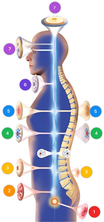
| Mode setting: 7 (Immunotherapy) Location: 1 (Sacrum) Morning: 15 minutes, Evening: 15 minutes |
Mode setting: 7 (Immunotherapy) Location: 1 (Sacrum) Morning: 30 minutes, Lunch: 30 minutes, Evening: 30 minutes |
Mode setting: 7 (Immunotherapy) Location: 1 (Sacrum) Morning: 30 minutes, Lunch: 30 minutes, Evening: 30 minutes |
| Total Morning: 60 minutes approx. $10 USD, Evening: 60 minutes approx. $10 USD |
Total Morning: 120 minutes approx. $20 USD, Lunch: 120 minutes approx. $20 USD, Evening: 120 minutes approx. $20 USD, |
Total Morning: 120 minutes approx. $20 USD, Lunch: 120 minutes approx. $20 USD, Evening: 120 minutes approx. $20 USD, |
| Usual treatment for 7-60 days approx. $140 USD – $1200 USD | Usual treatment for 6-8 weeks approx. $2,520 USD – $3,360 USD |
Usual treatment for 3-6 months approx. $5,400 USD – $10,800 USD
|
 |
|
Use the Mirari Cold Plasma device to treat Fibromyoma uterus effectively.
WARNING: MIRARI COLD PLASMA IS DESIGNED FOR THE HUMAN BODY WITHOUT ANY ARTIFICIAL OR THIRD PARTY PRODUCTS. USE OF OTHER PRODUCTS IN COMBINATION WITH MIRARI COLD PLASMA MAY CAUSE UNPREDICTABLE EFFECTS, HARM OR INJURY. PLEASE CONSULT A MEDICAL PROFESSIONAL BEFORE COMBINING ANY OTHER PRODUCTS WITH USE OF MIRARI.
Step 1: Cleanse the Skin
- Start by cleaning the affected area of the skin with a gentle cleanser or mild soap and water. Gently pat the area dry with a clean towel.
Step 2: Prepare the Mirari Cold Plasma device
- Ensure that the Mirari Cold Plasma device is fully charged or has fresh batteries as per the manufacturer’s instructions. Make sure the device is clean and in good working condition.
- Switch on the Mirari device using the power button or by following the specific instructions provided with the device.
- Some Mirari devices may have adjustable settings for intensity or treatment duration. Follow the manufacturer’s instructions to select the appropriate settings based on your needs and the recommended guidelines.
Step 3: Apply the Device
- Place the Mirari device in direct contact with the affected area of the skin. Gently glide or hold the device over the skin surface, ensuring even coverage of the area experiencing.
- Slowly move the Mirari device in a circular motion or follow a specific pattern as indicated in the user manual. This helps ensure thorough treatment coverage.
Step 4: Monitor and Assess:
- Keep track of your progress and evaluate the effectiveness of the Mirari device in managing your Fibromyoma uterus. If you have any concerns or notice any adverse reactions, consult with your health care professional.
Note
This guide is for informational purposes only and should not replace the advice of a medical professional. Always consult with your healthcare provider or a qualified medical professional for personal advice, diagnosis, or treatment. Do not solely rely on the information presented here for decisions about your health. Use of this information is at your own risk. The authors of this guide, nor any associated entities or platforms, are not responsible for any potential adverse effects or outcomes based on the content.
Mirari Cold Plasma System Disclaimer
- Purpose: The Mirari Cold Plasma System is a Class 2 medical device designed for use by trained healthcare professionals. It is registered for use in Thailand and Vietnam. It is not intended for use outside of these locations.
- Informational Use: The content and information provided with the device are for educational and informational purposes only. They are not a substitute for professional medical advice or care.
- Variable Outcomes: While the device is approved for specific uses, individual outcomes can differ. We do not assert or guarantee specific medical outcomes.
- Consultation: Prior to utilizing the device or making decisions based on its content, it is essential to consult with a Certified Mirari Tele-Therapist and your medical healthcare provider regarding specific protocols.
- Liability: By using this device, users are acknowledging and accepting all potential risks. Neither the manufacturer nor the distributor will be held accountable for any adverse reactions, injuries, or damages stemming from its use.
- Geographical Availability: This device has received approval for designated purposes by the Thai and Vietnam FDA. As of now, outside of Thailand and Vietnam, the Mirari Cold Plasma System is not available for purchase or use.
References
- Stewart, E. A., Cookson, C. L., Gandolfo, R. A., & Schulze-Rath, R. (2017). Epidemiology of uterine fibroids: a systematic review. BJOG: An International Journal of Obstetrics & Gynaecology, 124(10), 1501-1512. https://doi.org/10.1111/1471-0528.14640
- Zimmermann, A., Bernuit, D., Gerlinger, C., Schaefers, M., & Geppert, K. (2012). Prevalence, symptoms and management of uterine fibroids: an international internet-based survey of 21,746 women. BMC Women’s Health, 12, 6. https://doi.org/10.1186/1472-6874-12-6
- World Health Organization. (2020). International Classification of Primary Care, Second edition (ICPC-2). https://www.who.int/standards/classifications/other-classifications/international-classification-of-primary-care
- World Health Organization. (2019). International Statistical Classification of Diseases and Related Health Problems (ICD-10). https://icd.who.int/browse10/2019/en
- Munro, M. G., Critchley, H. O., & Fraser, I. S. (2011). The FIGO classification of causes of abnormal uterine bleeding in the reproductive years. Fertility and Sterility, 95(7), 2204-2208. https://doi.org/10.1016/j.fertnstert.2011.03.079
- Doherty, L., Mutlu, L., Sinclair, D., & Taylor, H. (2014). Uterine fibroids: clinical manifestations and contemporary management. Reproductive Sciences, 21(9), 1067-1092. https://doi.org/10.1177/1933719114533728
- Pritts, E. A., Parker, W. H., & Olive, D. L. (2009). Fibroids and infertility: an updated systematic review of the evidence. Fertility and Sterility, 91(4), 1215-1223. https://doi.org/10.1016/j.fertnstert.2008.01.051
- Bulun, S. E. (2013). Uterine fibroids. New England Journal of Medicine, 369(14), 1344-1355. https://doi.org/10.1056/NEJMra1209993
- Okolo, S. (2008). Incidence, aetiology and epidemiology of uterine fibroids. Best Practice & Research Clinical Obstetrics & Gynaecology, 22(4), 571-588. https://doi.org/10.1016/j.bpobgyn.2008.04.002
- Ciarmela, P., Islam, M. S., Reis, F. M., Gray, P. C., Bloise, E., Petraglia, F., Vale, W., & Castellucci, M. (2011). Growth factors and myometrium: biological effects in uterine fibroid and possible clinical implications. Human Reproduction Update, 17(6), 772-790. https://doi.org/10.1093/humupd/dmr031
- Wise, L. A., & Laughlin-Tommaso, S. K. (2016). Epidemiology of uterine fibroids: from menarche to menopause. Clinical Obstetrics and Gynecology, 59(1), 2-24. https://doi.org/10.1097/GRF.0000000000000164
- Stewart, E. A., Laughlin-Tommaso, S. K., Catherino, W. H., Lalitkumar, S., Gupta, D., & Vollenhoven, B. (2016). Uterine fibroids. Nature Reviews Disease Primers, 2, 16043. https://doi.org/10.1038/nrdp.2016.43
- Wise, L. A., & Laughlin-Tommaso, S. K. (2016). Epidemiology of uterine fibroids: from menarche to menopause. Clinical Obstetrics and Gynecology, 59(1), 2-24. https://doi.org/10.1097/GRF.0000000000000164
- Zimmermann, A., Bernuit, D., Gerlinger, C., Schaefers, M., & Geppert, K. (2012). Prevalence, symptoms and management of uterine fibroids: an international internet-based survey of 21,746 women. BMC Women’s Health, 12, 6. https://doi.org/10.1186/1472-6874-12-6
- Laughlin-Tommaso, S. K. (2018). Alternatives to hysterectomy: management of uterine fibroids. Obstetrics and Gynecology Clinics of North America, 45(4), 647-658. https://doi.org/10.1016/j.ogc.2016.04.001
- Munro, M. G., Critchley, H. O., & Fraser, I. S. (2011). The FIGO classification of causes of abnormal uterine bleeding in the reproductive years. Fertility and Sterility, 95(7), 2204-2208. https://doi.org/10.1016/j.fertnstert.2011.03.079
- Poppe, K., & Velkeniers, B. (2004). Thyroid disorders in infertile women. Annals of Endocrinology, 65(1), 93-101. https://pubmed.ncbi.nlm.nih.gov/12707633/
- Practice Committee of the American Society for Reproductive Medicine. (2015). Diagnostic evaluation of the infertile female: a committee opinion. Fertility and Sterility, 103(6), e44-e50. https://doi.org/10.1016/j.fertnstert.2015.03.019
- Kaump, G. R., & Spies, J. B. (2013). The impact of uterine artery embolization on ovarian function. Journal of Vascular and Interventional Radiology, 24(4), 459-467. https://doi.org/10.1016/j.jvir.2012.12.002
- Dueholm, M., Lundorf, E., Hansen, E. S., Ledertoug, S., & Olesen, F. (2002). Accuracy of magnetic resonance imaging and transvaginal ultrasonography in the diagnosis, mapping, and measurement of uterine myomas. American Journal of Obstetrics and Gynecology, 186(3), 409-415. https://doi.org/10.1067/mob.2002.121725
- Becker, E., Lev-Toaff, A. S., Kaufman, E. P., Halpern, E. J., Edelweiss, M. I., & Kurtz, A. B. (2002). The added value of transvaginal sonohysterography over transvaginal sonography alone in women with known or suspected leiomyoma. Journal of Ultrasound in Medicine, 21(3), 237-247. https://doi.org/10.7863/jum.2002.21.3.237
- Clark, T. J., & Gupta, J. K. (2002). Endometrial sampling of gynaecological pathology. The Obstetrician & Gynaecologist, 4(3), 169-174. https://doi.org/10.1576/toag.2002.4.3.169
- Di Spiezio Sardo, A., Mazzon, I., Bramante, S., Bettocchi, S., Bifulco, G., Guida, M., & Nappi, C. (2008). Hysteroscopic myomectomy: a comprehensive review of surgical techniques. Human Reproduction Update, 14(2), 101-119. https://doi.org/10.1093/humupd/dmm041
- Mehine, M., Mäkinen, N., Heinonen, H. R., Aaltonen, L. A., & Vahteristo, P. (2014). Genomics of uterine leiomyomas: insights from high-throughput sequencing. Fertility and Sterility, 102(3), 621-629. https://doi.org/10.1016/j.fertnstert.2014.06.050
- Laughlin-Tommaso, S. K., Hesley, G. K., Hopkins, M. R., Brandt, K. R., Zhu, Y., & Stewart, E. A. (2017). Clinical limitations of the International Federation of Gynecology and Obstetrics (FIGO) classification of uterine fibroids. International Journal of Gynecology & Obstetrics, 139(2), 143-148. https://doi.org/10.1002/ijgo.12266
- Vilos, G. A., Allaire, C., Laberge, P. Y., Leyland, N., Special Contributors, Vilos, A. G., … & Chen, I. (2015). The management of uterine leiomyomas. Journal of Obstetrics and Gynaecology Canada, 37(2), 157-178. https://doi.org/10.1016/S1701-2163(15)30338-8
- Ghant, M. S., Sengoba, K. S., Recht, H., Cameron, K. A., Lawson, A. K., & Marsh, E. E. (2015). Beyond the physical: a qualitative assessment of the burden of symptomatic uterine fibroids on women’s emotional and psychosocial health. Journal of Psychosomatic Research, 78(5), 499-503. https://doi.org/10.1016/j.jpsychores.2014.12.016
- Lethaby, A., Vollenhoven, B., & Sowter, M. (2002). Pre-operative GnRH analogue therapy before hysterectomy or myomectomy for uterine fibroids. Cochrane Database of Systematic Reviews, (2), CD000547. https://doi.org/10.1002/14651858.CD000547
- Donnez, J., Tatarchuk, T. F., Bouchard, P., Puscasiu, L., Zakharenko, N. F., Ivanova, T., … & Østergaard, A. (2012). Ulipristal acetate versus placebo for fibroid treatment before surgery. New England Journal of Medicine, 366(5), 409-420. https://doi.org/10.1056/NEJMoa1103182
- Lethaby, A., Duckitt, K., & Farquhar, C. (2013). Non‐steroidal anti‐inflammatory drugs for heavy menstrual bleeding. Cochrane Database of Systematic Reviews, (1), CD000400. https://doi.org/10.1002/14651858.CD000400.pub3
- Naoulou, B., & Tsai, M. C. (2012). Efficacy of tranexamic acid in the treatment of idiopathic and non-functional heavy menstrual bleeding: a systematic review. Acta Obstetricia et Gynecologica Scandinavica, 91(5), 529-537. https://doi.org/10.1111/j.1600-0412.2012.01361.x
- de Bastos, M., Stegeman, B. H., Rosendaal, F. R., Van Hylckama Vlieg, A., Helmerhorst, F. M., Stijnen, T., & Dekkers, O. M. (2014). Combined oral contraceptives: venous thrombosis. Cochrane Database of Systematic Reviews, (3), CD010813. https://doi.org/10.1002/14651858.CD010813.pub2
- Parsanezhad, M. E., Azmoon, M., Alborzi, S., Rajaeefard, A., Zarei, A., Kazerooni, T., … & Schmidt, E. H. (2010). A randomized, controlled clinical trial comparing the effects of aromatase inhibitor (letrozole) and gonadotropin-releasing hormone agonist (triptorelin) on uterine leiomyoma volume and hormonal status. Fertility and sterility, 93(1), 192-198. https://doi.org/10.1016/j.fertnstert.2008.09.064
- De Leo, V., Morgante, G., La Marca, A., Musacchio, M. C., Sorace, M., Cavicchioli, C., & Petraglia, F. (2002). A benefit-risk assessment of medical treatment for uterine leiomyomas. Drug safety, 25(11), 759-779. https://doi.org/10.2165/00002018-200225110-00003
- Engman, M., Granberg, S., Williams, A. R., Meng, C. X., Lalitkumar, P. G., & Gemzell-Danielsson, K. (2009). Mifepristone for treatment of uterine leiomyoma. A prospective randomized placebo controlled trial. Human reproduction, 24(8), 1870-1879. https://doi.org/10.1093/humrep/dep100
- Cardozo, E. R., Clark, A. D., Banks, N. K., Henne, M. B., Stegmann, B. J., & Segars, J. H. (2012). The estimated annual cost of uterine leiomyomata in the United States. American journal of obstetrics and gynecology, 206(3), 211-e1. https://doi.org/10.1016/j.ajog.2011.12.002
- Chen, N. N., Han, M., Yang, H., Yang, G. Y., Wang, Y. Y., Wu, X. K., & Liu, J. P. (2014). Chinese herbal medicine Guizhi Fuling Formula for treatment of uterine fibroids: a systematic review of randomised clinical trials. BMC complementary and alternative medicine, 14(1), 2. https://doi.org/10.1186/1472-6882-14-2
- Liu, J. P., Yang, H., Xia, Y., & Cardini, F. (2013). Herbal preparations for uterine fibroids. Cochrane Database of Systematic Reviews, (4). https://doi.org/10.1002/14651858.CD005292.pub3
- Yang, J. H., Chen, C. D., Chen, S. U., Yang, Y. S., & Chen, M. J. (2018). The impact of submucous myoma on reproductive outcomes in patients with intramural myomas undergoing IVF–ICSI. Journal of Assisted Reproduction and Genetics, 35(12), 2167-2174. https://doi.org/10.1007/s10815-018-1301-7
- Chiaffarino, F., Parazzini, F., La Vecchia, C., Chatenoud, L., Di Cintio, E., & Marsico, S. (1999). Diet and uterine myomas. Obstetrics & Gynecology, 94(3), 395-398. https://doi.org/10.1016/s0029-7844(99)00305-1
- Stein, A., & Mallozzi, S. S. (2006). Pelvic floor muscle training for pelvic organ prolapse. Physical Therapy Reviews, 11(3), 169-175. https://doi.org/10.1179/108331906X144064
- Wise, L. A., Palmer, J. R., Spiegelman, D., Harlow, B. L., Stewart, E. A., Adams-Campbell, L. L., & Rosenberg, L. (2005). Influence of body size and body fat distribution on risk of uterine leiomyomata in US black women. Epidemiology, 16(3), 346-354. https://doi.org/10.1097/01.ede.0000158742.11877.99
- Baird, D. D., Dunson, D. B., Hill, M. C., Cousins, D., & Schectman, J. M. (2007). Association of physical activity with development of uterine leiomyoma. American journal of epidemiology, 165(2), 157-163. https://doi.org/10.1093/aje/kwj363
- Vines, A. I., Ta, M., & Esserman, D. A. (2010). The association between self-reported major life events and the presence of uterine fibroids. Women’s Health Issues, 20(4), 294-298. https://doi.org/10.1016/j.whi.2010.03.009
- Wise, L. A., Laughlin-Tommaso, S. K., Rubin, S., Wesselink, A., Hahn, K. A., Mikkelsen, E. M., … & Rothman, K. J. (2020). Prenatal and perinatal factors and risk of uterine leiomyomata. Annals of epidemiology, 41, 1-7. https://doi.org/10.1016/j.annepidem.2019.12.008
- Brakta, S., Diamond, J. S., Al-Hendy, A., Diamond, M. P., & Halder, S. K. (2015). Role of vitamin D in uterine fibroid biology. Fertility and sterility, 104(3), 698-706. https://doi.org/10.1016/j.fertnstert.2015.05.031
Related articles
Made in USA


