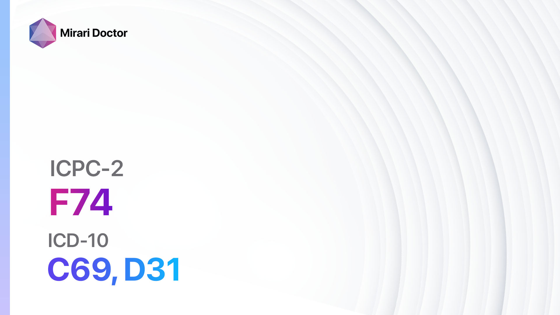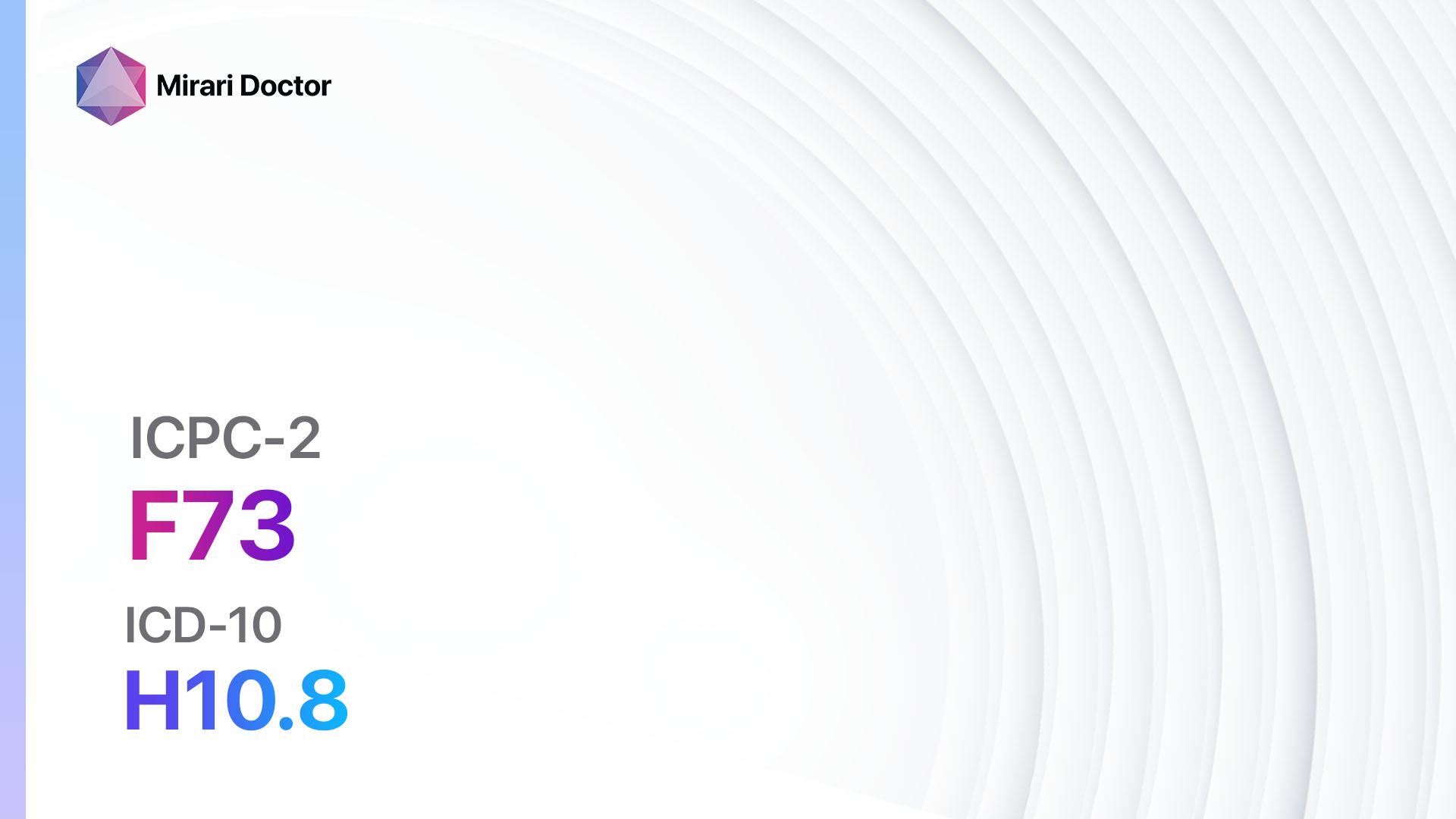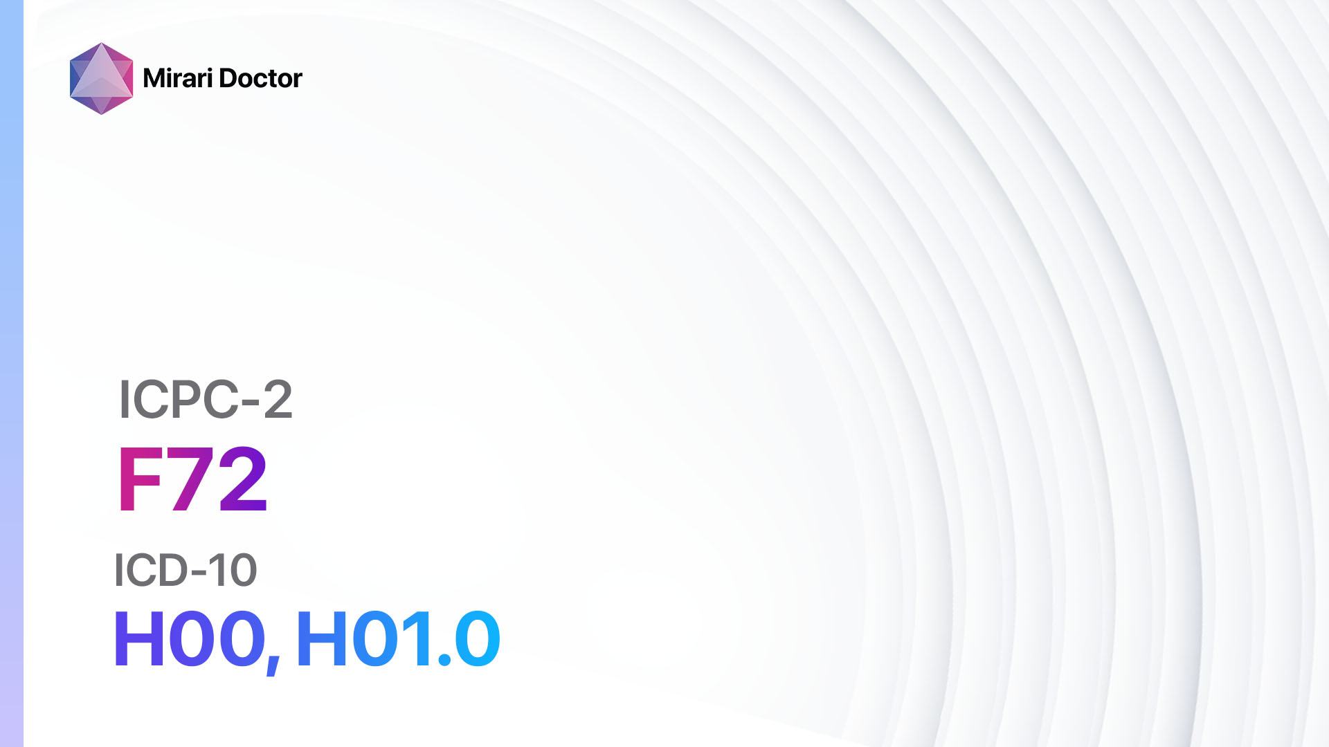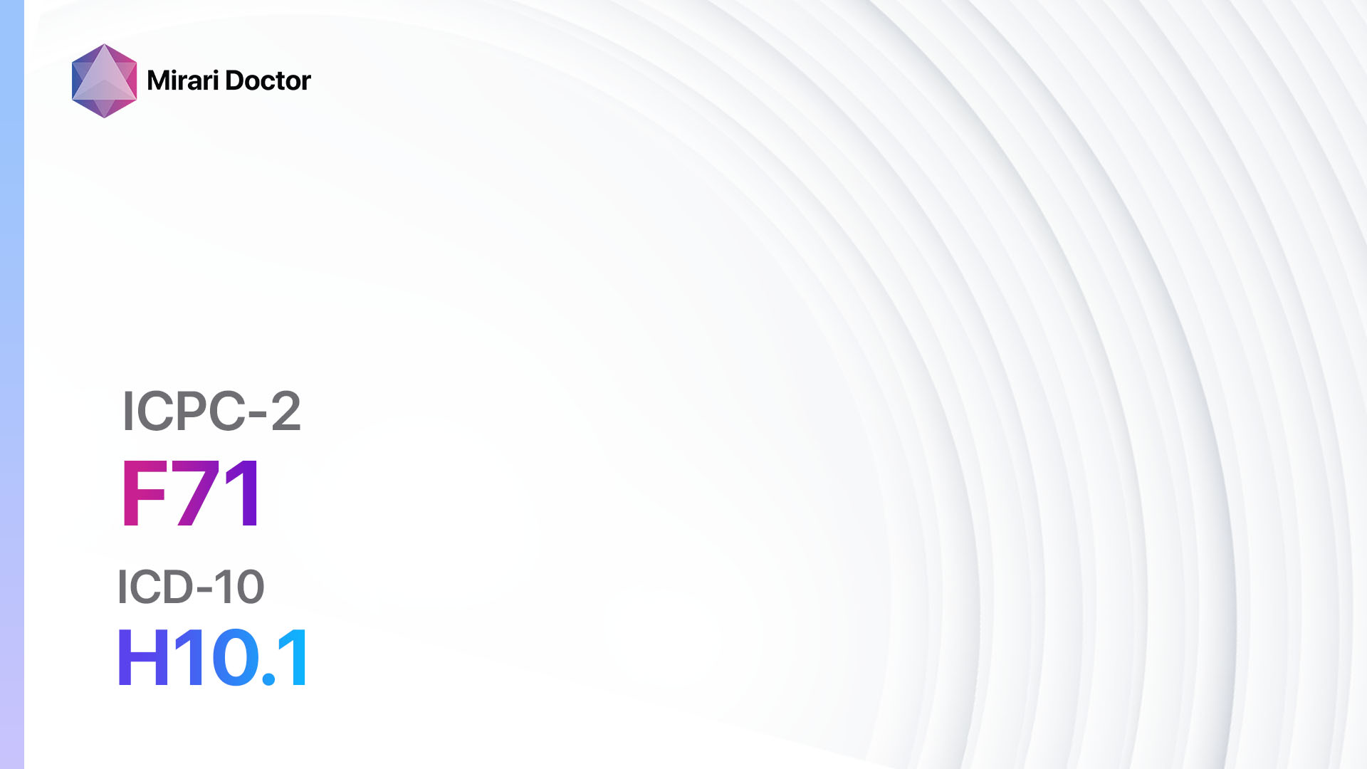
Introduction
Corneal ulcer is a serious condition characterized by an open sore on the cornea, the clear front surface of the eye. It can lead to vision loss if not promptly diagnosed and treated.[1] The aim of this guide is to provide healthcare professionals with a comprehensive overview of the symptoms, causes, diagnostic steps, possible interventions, and patient education related to corneal ulcers.
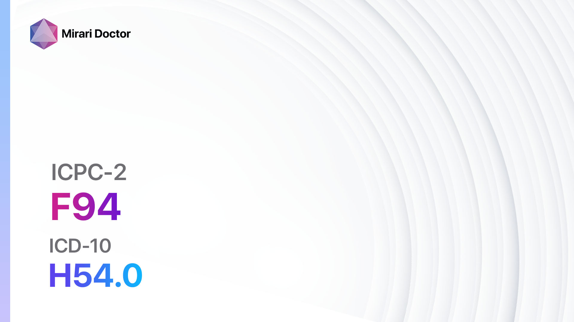
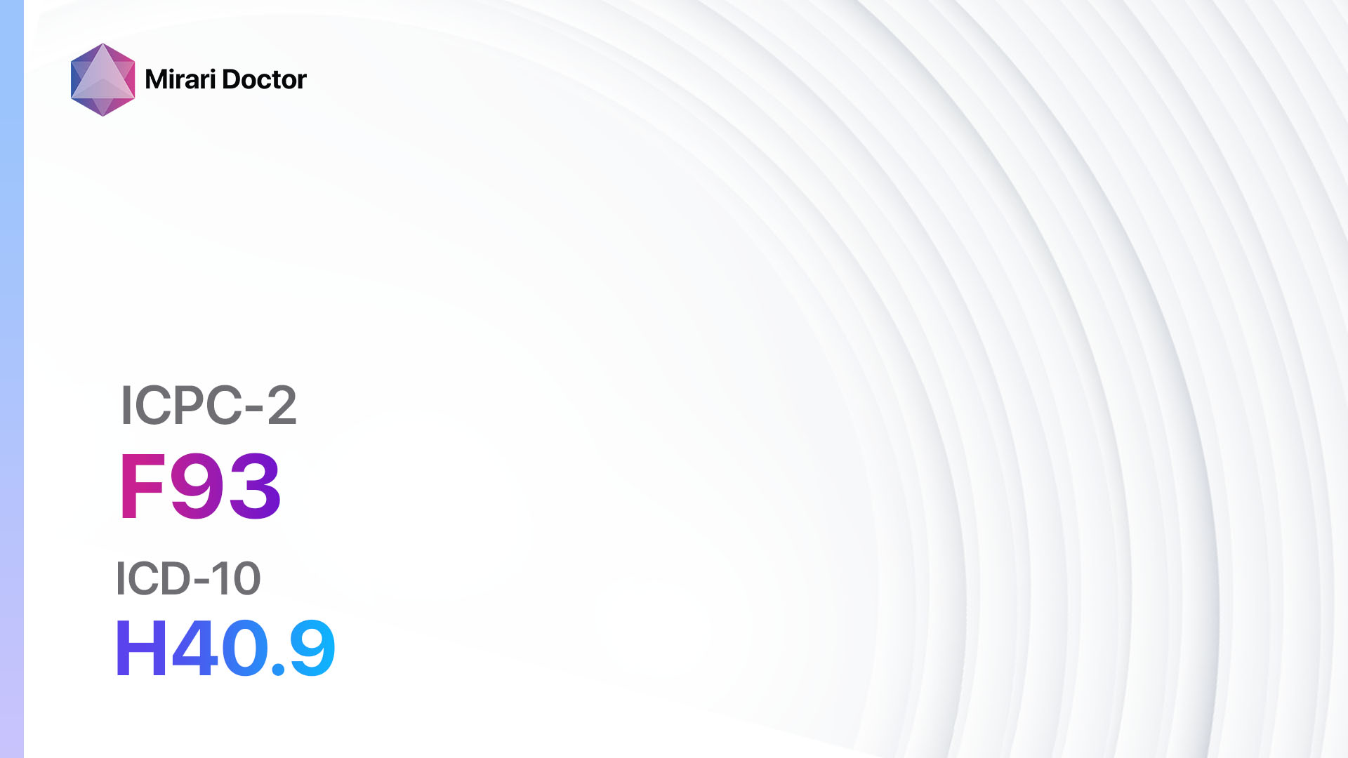
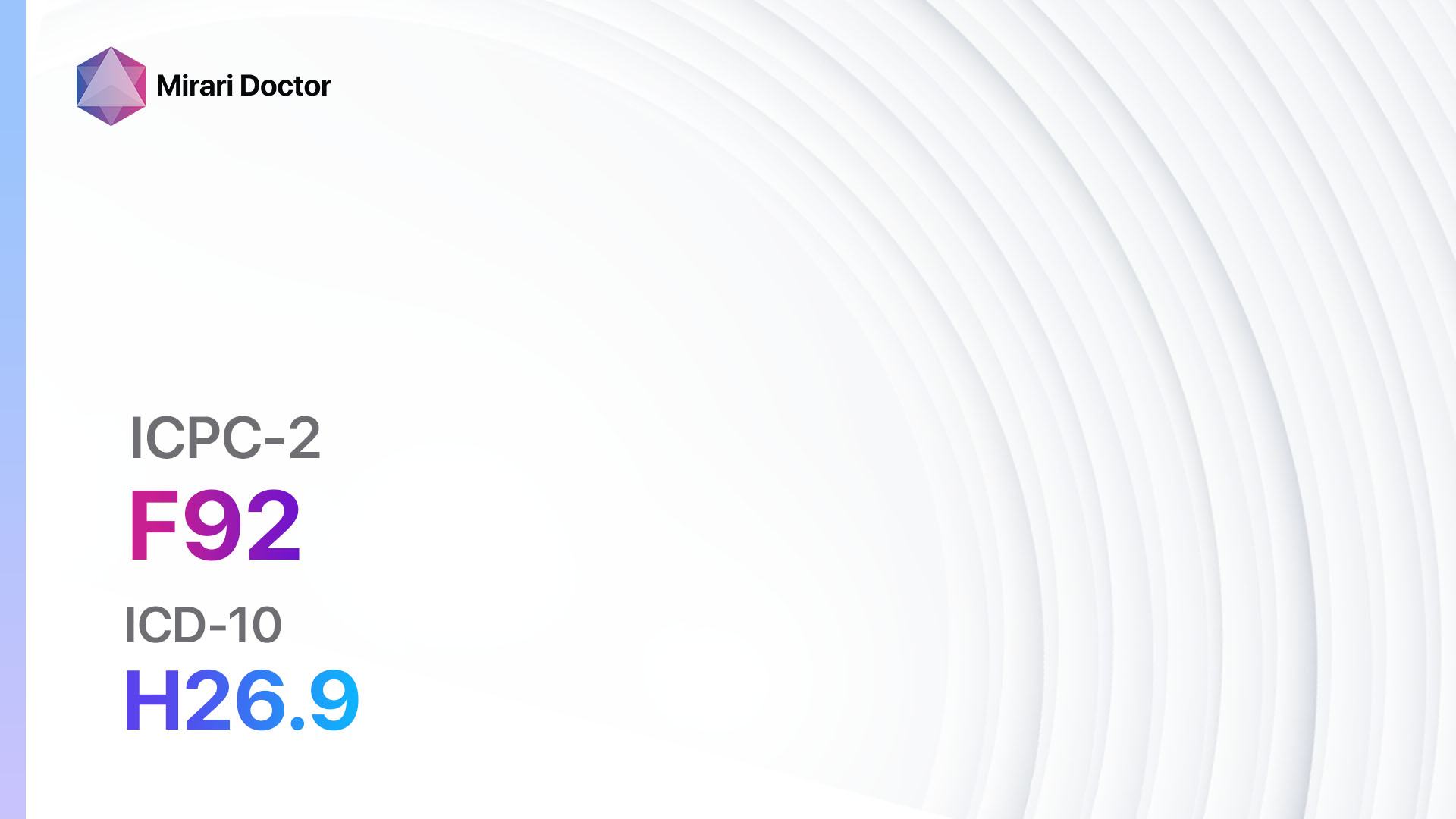
Codes
Symptoms
- Eye redness: The affected eye may appear red or bloodshot.[4]
- Eye pain: Patients may experience moderate to severe pain in the affected eye.[4]
- Blurred vision: Vision may become blurry or hazy.[4]
- Sensitivity to light: Patients may experience increased sensitivity to light.[4]
- Excessive tearing: The affected eye may produce excessive tears.[4]
- Eye discharge: Patients may have a yellowish or greenish discharge from the affected eye.[4]
Causes
- Bacterial infection: Bacteria, such as Staphylococcus aureus or Pseudomonas aeruginosa, can cause corneal ulcers.[5]
- Viral infection: Viruses, such as herpes simplex virus or varicella-zoster virus, can lead to corneal ulcers.[5]
- Fungal infection: Fungi, such as Candida or Aspergillus, can cause corneal ulcers.[5]
- Trauma: Injury to the cornea, such as a scratch or foreign body, can result in corneal ulcers.[6]
- Contact lens wear: Improper use or poor hygiene of contact lenses can increase the risk of corneal ulcers.[6]
Diagnostic Steps
Medical History
- Gather information about the patient’s risk factors, such as contact lens use, recent eye trauma, or previous eye infections.[7]
- Inquire about symptoms, including eye pain, redness, blurred vision, and sensitivity to light.[7]
- Ask about any underlying medical conditions, such as diabetes or immunosuppression, which may increase the risk of corneal ulcers.[7]
Physical Examination
- Inspect the affected eye for redness, swelling, discharge, or any visible corneal abnormalities.[8]
- Assess visual acuity to determine the extent of vision loss.[8]
- Perform a slit-lamp examination to examine the cornea in detail and identify any corneal ulcers or infiltrates.[8]
Laboratory Tests
- Corneal scraping: Collect a sample of the corneal tissue for microbiological analysis to identify the causative organism.[9]
- Culture and sensitivity testing: Perform a culture of the corneal scraping to determine the specific bacteria, virus, or fungus causing the corneal ulcer and assess its sensitivity to various antimicrobial agents.[9]
Diagnostic Imaging
- Fluorescein staining: Apply fluorescein dye to the eye to identify and visualize corneal ulcers or defects.[8]
- Anterior segment optical coherence tomography (OCT): Use OCT imaging to assess the depth and extent of corneal ulcers and evaluate the involvement of underlying structures.[8]
Other Tests
- Intraocular pressure measurement: Measure the intraocular pressure to rule out glaucoma, which can be associated with corneal ulcers.[8]
- Blood tests: Perform blood tests, such as complete blood count and inflammatory markers, to assess the overall health status and identify any underlying systemic conditions.[8]
Follow-up and Patient Education
- Schedule regular follow-up appointments to monitor the progress of the corneal ulcer and adjust treatment if necessary.[10]
- Educate the patient about the importance of proper contact lens hygiene and the risks associated with wearing contact lenses.[10]
- Instruct the patient to avoid rubbing or touching the affected eye and to follow the prescribed treatment regimen diligently.[10]
- Emphasize the need for prompt medical attention if symptoms worsen or if new symptoms, such as severe eye pain or vision loss, develop.[10]
Possible Interventions
Traditional Interventions
Medications:
Top 5 drugs for Corneal Ulcer:
- Fluoroquinolone eye drops (e.g., Ciprofloxacin, Ofloxacin):
- Cost: $10-$50 per bottle.
- Contraindications: Hypersensitivity to fluoroquinolones.
- Side effects: Eye irritation, burning sensation.
- Severe side effects: Corneal perforation, allergic reactions.
- Drug interactions: None reported.
- Warning: Use with caution in patients with a history of tendon disorders.
- Broad-spectrum antibiotic eye drops (e.g., Tobramycin, Gentamicin):
- Cost: $10-$50 per bottle.
- Contraindications: Hypersensitivity to aminoglycosides.
- Side effects: Eye irritation, stinging.
- Severe side effects: Corneal toxicity, allergic reactions.
- Drug interactions: None reported.
- Warning: Prolonged use may increase the risk of fungal or resistant bacterial infections.
- Antiviral eye drops (e.g., Trifluridine, Ganciclovir):
- Cost: $50-$100 per bottle.
- Contraindications: Hypersensitivity to antiviral agents.
- Side effects: Eye irritation, stinging.
- Severe side effects: Corneal toxicity, allergic reactions.
- Drug interactions: None reported.
- Warning: Use with caution in patients with compromised corneal integrity.
- Antifungal eye drops (e.g., Natamycin, Amphotericin B):
- Cost: $50-$100 per bottle.
- Contraindications: Hypersensitivity to antifungal agents.
- Side effects: Eye irritation, burning sensation.
- Severe side effects: Corneal toxicity, allergic reactions.
- Drug interactions: None reported.
- Warning: Use with caution in patients with compromised corneal integrity.
- Corticosteroid eye drops (e.g., Prednisolone, Dexamethasone):
- Cost: $10-$50 per bottle.
- Contraindications: Active viral or fungal infections.
- Side effects: Increased intraocular pressure, cataract formation.
- Severe side effects: Corneal thinning, delayed wound healing.
- Drug interactions: None reported.
- Warning: Use with caution and under close supervision due to the risk of exacerbating infection or delaying healing.
Surgical Procedures:
- Corneal debridement: Removal of necrotic tissue and debris from the corneal ulcer to promote healing. Cost: $500-$1,500.
- Corneal patch graft: Placement of a donor corneal tissue over the ulcer to facilitate healing and restore corneal integrity. Cost: $2,000-$5,000.
- Amniotic membrane transplantation: Application of amniotic membrane to the corneal ulcer to promote healing and reduce inflammation. Cost: $3,000-$6,000.
Alternative Interventions
- Autologous serum eye drops: Eye drops made from the patient’s own blood serum, which contain growth factors and anti-inflammatory substances. Cost: $100-$200 per bottle.
- Bandage contact lens: Placement of a therapeutic contact lens over the corneal ulcer to protect the cornea and promote healing. Cost: $100-$300 per lens.
- Amniotic membrane extract eye drops: Eye drops containing amniotic membrane extract, which has anti-inflammatory and wound healing properties. Cost: $200-$400 per bottle.
- Platelet-rich plasma eye drops: Eye drops made from the patient’s own platelet-rich plasma, which contains growth factors that promote healing. Cost: $300-$500 per bottle.
- Stem cell therapy: Injection of stem cells into the corneal ulcer to stimulate tissue regeneration and healing. Cost: $5,000-$10,000 per treatment.
Lifestyle Interventions
- Eye hygiene: Instruct the patient to maintain good eye hygiene by washing hands before touching the eyes and avoiding rubbing or scratching the affected eye.
- Contact lens care: Educate the patient about proper contact lens care, including regular cleaning, disinfection, and replacement as recommended by the manufacturer.
- Eye protection: Advise the patient to wear protective eyewear, such as goggles, when engaging in activities that may pose a risk of eye injury.
- Healthy lifestyle: Encourage the patient to maintain a healthy lifestyle, including a balanced diet, regular exercise, and adequate sleep, to support overall eye health.
- Avoidance of irritants: Advise the patient to avoid exposure to irritants, such as smoke, dust, and chemicals, which can exacerbate symptoms and delay healing.
It is important to note that the cost ranges provided are approximate and may vary depending on the location and availability of the interventions.
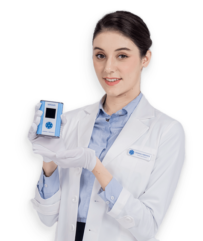
Mirari Cold Plasma Alternative Intervention
Understanding Mirari Cold Plasma
- Safe and Non-Invasive Treatment: Mirari Cold Plasma is a safe and non-invasive treatment option for various skin conditions. It does not require incisions, minimizing the risk of scarring, bleeding, or tissue damage.
- Efficient Extraction of Foreign Bodies: Mirari Cold Plasma facilitates the removal of foreign bodies from the skin by degrading and dissociating organic matter, allowing easier access and extraction.
- Pain Reduction and Comfort: Mirari Cold Plasma has a local analgesic effect, providing pain relief during the treatment, making it more comfortable for the patient.
- Reduced Risk of Infection: Mirari Cold Plasma has antimicrobial properties, effectively killing bacteria and reducing the risk of infection.
- Accelerated Healing and Minimal Scarring: Mirari Cold Plasma stimulates wound healing and tissue regeneration, reducing healing time and minimizing the formation of scars.
Mirari Cold Plasma Prescription
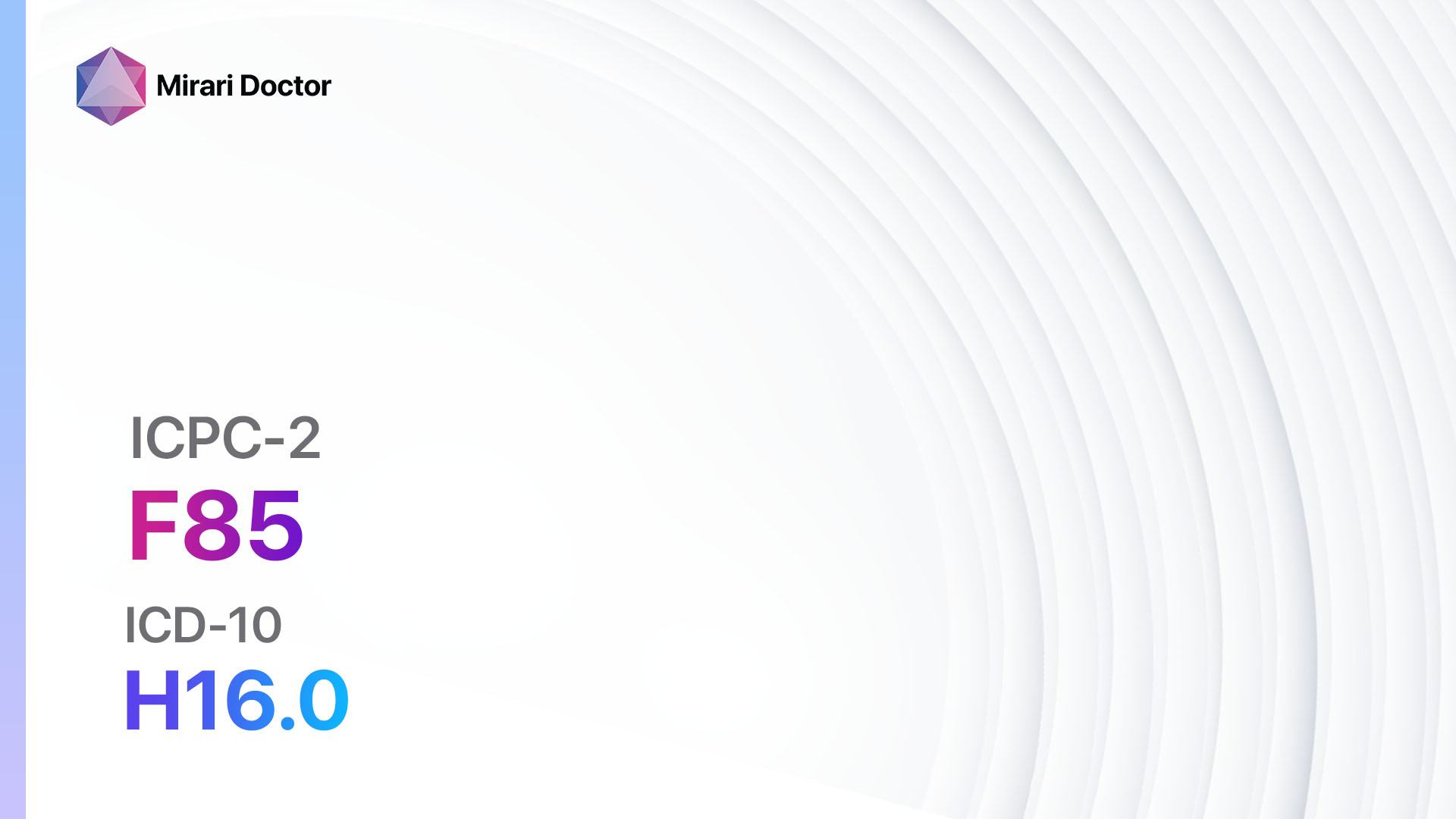
Video instructions for using Mirari Cold Plasma Device – F85 Corneal ulcer (ICD-10:H16.0)
| Mild | Moderate | Severe |
| Mode setting: 1 (Infection) Location: 7 (Neuro system & ENT) Morning: 15 minutes, Evening: 15 minutes |
Mode setting: 1 (Infection) Location: 7 (Neuro system & ENT) Morning: 30 minutes, Lunch: 30 minutes, Evening: 30 minutes |
Mode setting: 1 (Infection) Location: 7 (Neuro system & ENT) Morning: 30 minutes, Lunch: 30 minutes, Evening: 30 minutes |
| Mode setting: 2 (Wound Healing) Location: 7 (Neuro system & ENT) Morning: 15 minutes, Evening: 15 minutes |
Mode setting: 2 (Wound Healing) Location: 7 (Neuro system & ENT) Morning: 30 minutes, Lunch: 30 minutes, Evening: 30 minutes |
Mode setting: 2 (Wound Healing) Location: 7 (Neuro system & ENT) Morning: 30 minutes, Lunch: 30 minutes, Evening: 30 minutes |
| Mode setting: 3 (Antiviral Therapy) Location: 7 (Neuro system & ENT) Morning: 15 minutes, Evening: 15 minutes |
Mode setting: 3 (Antiviral Therapy) Location: 7 (Neuro system & ENT) Morning: 30 minutes, Lunch: 30 minutes, Evening: 30 minutes |
Mode setting: 3 (Antiviral Therapy) Location: 7 (Neuro system & ENT) Morning: 30 minutes, Lunch: 30 minutes, Evening: 30 minutes |
| Total Morning: 45 minutes approx. $7.50 USD, Evening: 45 minutes approx. $7.50 USD |
Total Morning: 90 minutes approx. $15 USD, Lunch: 90 minutes approx. $15 USD, Evening: 90 minutes approx. $15 USD, |
Total Morning: 90 minutes approx. $15 USD, Lunch: 90 minutes approx. $15 USD, Evening: 90 minutes approx. $15 USD, |
| Usual treatment for 7-60 days approx. $105 USD – $900 USD | Usual treatment for 6-8 weeks approx. $1,890 USD – $2,520 USD |
Usual treatment for 3-6 months approx. $4,050 USD – $8,100 USD
|
 |
|
Use the Mirari Cold Plasma device to treat Corneal ulcer effectively.
WARNING: MIRARI COLD PLASMA IS DESIGNED FOR THE HUMAN BODY WITHOUT ANY ARTIFICIAL OR THIRD PARTY PRODUCTS. USE OF OTHER PRODUCTS IN COMBINATION WITH MIRARI COLD PLASMA MAY CAUSE UNPREDICTABLE EFFECTS, HARM OR INJURY. PLEASE CONSULT A MEDICAL PROFESSIONAL BEFORE COMBINING ANY OTHER PRODUCTS WITH USE OF MIRARI.
Step 1: Cleanse the Skin
- Start by cleaning the affected area of the skin with a gentle cleanser or mild soap and water. Gently pat the area dry with a clean towel.
Step 2: Prepare the Mirari Cold Plasma device
- Ensure that the Mirari Cold Plasma device is fully charged or has fresh batteries as per the manufacturer’s instructions. Make sure the device is clean and in good working condition.
- Switch on the Mirari device using the power button or by following the specific instructions provided with the device.
- Some Mirari devices may have adjustable settings for intensity or treatment duration. Follow the manufacturer’s instructions to select the appropriate settings based on your needs and the recommended guidelines.
Step 3: Apply the Device
- Place the Mirari device in direct contact with the affected area of the skin. Gently glide or hold the device over the skin surface, ensuring even coverage of the area experiencing.
- Slowly move the Mirari device in a circular motion or follow a specific pattern as indicated in the user manual. This helps ensure thorough treatment coverage.
Step 4: Monitor and Assess:
- Keep track of your progress and evaluate the effectiveness of the Mirari device in managing your Corneal ulcer. If you have any concerns or notice any adverse reactions, consult with your health care professional.
Note
This guide is for informational purposes only and should not replace the advice of a medical professional. Always consult with your healthcare provider or a qualified medical professional for personal advice, diagnosis, or treatment. Do not solely rely on the information presented here for decisions about your health. Use of this information is at your own risk. The authors of this guide, nor any associated entities or platforms, are not responsible for any potential adverse effects or outcomes based on the content.
Mirari Cold Plasma System Disclaimer
- Purpose: The Mirari Cold Plasma System is a Class 2 medical device designed for use by trained healthcare professionals. It is registered for use in Thailand and Vietnam. It is not intended for use outside of these locations.
- Informational Use: The content and information provided with the device are for educational and informational purposes only. They are not a substitute for professional medical advice or care.
- Variable Outcomes: While the device is approved for specific uses, individual outcomes can differ. We do not assert or guarantee specific medical outcomes.
- Consultation: Prior to utilizing the device or making decisions based on its content, it is essential to consult with a Certified Mirari Tele-Therapist and your medical healthcare provider regarding specific protocols.
- Liability: By using this device, users are acknowledging and accepting all potential risks. Neither the manufacturer nor the distributor will be held accountable for any adverse reactions, injuries, or damages stemming from its use.
- Geographical Availability: This device has received approval for designated purposes by the Thai and Vietnam FDA. As of now, outside of Thailand and Vietnam, the Mirari Cold Plasma System is not available for purchase or use.
References
- Eghrari AO, Riazuddin SA, Gottsch JD. Overview of the Cornea: Structure, Function, and Development. Prog Mol Biol Transl Sci. 2015;134:7-23. doi:10.1016/bs.pmbts.2015.04.001
- World Organization of Family Doctors (WONCA). International Classification of Primary Care, Second edition (ICPC-2). Oxford University Press; 1998.
- World Health Organization. International Statistical Classification of Diseases and Related Health Problems (ICD-10). World Health Organization; 2019.
- Farahani M, Patel R, Dwarakanathan S. Infectious corneal ulcers. Dis Mon. 2017;63(2):33-37. doi:10.1016/j.disamonth.2016.09.005
- Austin A, Lietman T, Rose-Nussbaumer J. Update on the Management of Infectious Keratitis. Ophthalmology. 2017;124(11):1678-1689. doi:10.1016/j.ophtha.2017.05.012
- Bourcier T, Thomas F, Borderie V, Chaumeil C, Laroche L. Bacterial keratitis: predisposing factors, clinical and microbiological review of 300 cases. Br J Ophthalmol. 2003;87(7):834-838. doi:10.1136/bjo.87.7.834
- Tananuvat N, Punyakhum O, Ausayakhun S, Chaidaroon W. Etiology and clinical outcomes of microbial keratitis at a tertiary eye-care center in northern Thailand. J Med Assoc Thai. 2012;95 Suppl 4:S8-S17.
- Dalmon C, Porco TC, Lietman TM, et al. The clinical differentiation of bacterial and fungal keratitis: a photographic survey. Invest Ophthalmol Vis Sci. 2012;53(4):1787-1791. doi:10.1167/iovs.11-8478
- Sharma S. Diagnosis of infectious diseases of the eye. Eye (Lond). 2012;26(2):177-184. doi:10.1038/eye.2011.275
- Ung L, Bispo PJM, Shanbhag SS, Gilmore MS, Chodosh J. The persistent dilemma of microbial keratitis: Global burden, diagnosis, and antimicrobial resistance. Surv Ophthalmol. 2019;64(3):255-271. doi:10.1016/j.survophthal.2018.12.003
Related articles
Made in USA
