
介绍
不属于特定类别的眼部症状或投诉可能难以诊断和治疗。本指南旨在为医护专业人员提供一种综合的方法,以评估和管理具有不属于特定诊断的眼部症状或投诉的患者。通过遵循本指南中列出的步骤,医护专业人员可以收集相关信息,进行必要的检查和测试,并为患者提供适当的干预措施。[1][2]
代码
- ICPC-2 代码: F29 其他眼症状/投诉[3]
- ICD-10 代码: H57.9 未指定的眼及附件疾病[4]
症状
- 视力模糊:患者可能抱怨视物不清或视力不锐利。[5]
- 眼痛:患者可能在一只或双眼中感到不适或疼痛。[6]
- 眼睛红肿:眼睛可能显得红或充血。[7]
- 瘙痒:患者可能报告眼睛发痒或有异物感。[8]
- 干涩:患者可能抱怨眼睛干燥或有砂砾感。[9]
- 光敏感:患者可能发现强光或阳光刺眼或不适。[10]
- 流泪过多:患者可能会有水汪汪的眼睛,且无明显原因。
- 飞蚊症:患者可能看到视野中有小斑点或斑痕漂浮。
- 光闪:患者可能看到短暂的闪光或光芒。
- 重影:患者可能看到两个影像,而不是一个。
- 眼部分泌物:患者的眼睛可能有分泌物,表现为水状、粘性或脓样。
原因
- 眼疲劳:长时间使用数字设备、阅读或其他需要集中注意力的活动会导致眼疲劳。
- 干眼症:泪液分泌不足或泪液质量差会导致眼干。
- 过敏:对花粉、灰尘或其他刺激物的过敏反应会引起眼部症状。
- 结膜炎:结膜的炎症,通常由于感染或过敏导致,可能导致红肿、瘙痒和分泌物。
- 异物:眼内有异物存在会导致刺激和不适。
- 睑缘炎:眼睑的炎症会导致红肿、瘙痒和结痂。
- 偏头痛:某些偏头痛会导致视觉障碍,如光闪或暂时失明。
- 屈光不正:未矫正的近视、远视或散光会导致视力模糊。
- 白内障:眼内晶状体混浊会导致视力模糊或减弱。
- 青光眼:眼压增加会损害视神经而导致视力丧失。
- 视网膜脱离:视网膜从眼后部脱离会导致飞蚊症、光闪和幕状阴影在视野中出现。
诊断步骤
病史采集
- 收集患者症状的信息,包括持续时间、严重程度以及任何相关因素(例如活动、时间)。
- 询问任何既往眼病或手术史。
- 询问可能导致眼部症状的任何病况或药物。
- 评估任何风险因素,如家族眼病史或接触刺激物的可能性。
体格检查
- 检查眼部外部结构,包括眼睑、结膜和巩膜,以发现任何异常。
- 采用视力表或其他适当的方法评估视力。
- 进行裂隙灯检查以评估眼前段,包括角膜、虹膜和晶状体。
- 散瞳检查眼后段,包括视网膜和视神经。
- 使用眼压计测量眼内压以筛查青光眼。
实验室检查
- 全血计数(CBC):评估可能导致眼部症状的潜在全身性病况。
- 过敏测试:如果怀疑过敏是眼部症状的原因,可以测试特定过敏原以识别触发因素。
- 泪膜评价:如泪液破裂时间(TBUT)和Schirmer试验,用于评估泪液分泌和质量,尤其在干眼症情况下。
诊断成像
- 光学相干断层扫描(OCT):这种非侵入性成像技术提供视网膜的详细横断面图像,有助于诊断黄斑变性或糖尿病视网膜病变等病况。
- 眼底摄影:可获得高分辨率视网膜图像以记录任何异常或随时间的变化。
- 荧光素血管造影:这种成像技术使用荧光染料以评估视网膜血流,有助于诊断视网膜血管阻塞或黄斑变性等病况。
其他检查
- 视野测试:评估患者的周边视觉,可以帮助检测青光眼或视神经损伤等病况。
- 角膜地形图:绘制角膜的曲率图,有助于诊断圆锥角膜或角膜营养不良。
- 视网膜电流图(ERG):测量视网膜对于光刺激的电反应,能帮助诊断视网膜疾病。
随访与患者教育
- 根据患者的病情和治疗计划确定随访预约。
- 提供正确的眼部护理建议,包括卫生、使用润滑性眼药水以及防护于刺激物或过敏原。
- 讨论生活方式调整,例如减少屏幕时间或戴护目镜,如适用。
- 解决患者对其眼部症状或治疗的任何疑虑或疑问。
可能的干预措施
传统干预
药物:
针对其他眼部症状/投诉的首选5种药物:
- 人工泪液:
- 费用:每瓶5至15美元。
- 禁忌症:对成分的过敏反应。
- 副作用:暂时性的视力模糊。
- 严重副作用:未有报道。
- 药物相互作用:未有报道。
- 警告:按指示使用并避免瓶子的污染。
- 抗组胺眼药水(例如,酮替芬):
- 费用:每瓶10至20美元。
- 禁忌症:对成分的过敏反应。
- 副作用:暂时性的刺痛或烧灼感。
- 严重副作用:未有报道。
- 药物相互作用:未有报道。
- 警告:按指示使用并避免与软性隐形眼镜接触。
- 局部皮质类固醇(例如,泼尼松龙):
- 费用:每管10至30美元。
- 禁忌症:活动性病毒、真菌或细菌眼感染。
- 副作用:眼内压增高、白内障形成。
- 严重副作用:短期使用未有报道。
- 药物相互作用:未有报道。
- 警告:需在眼科医生的监督下使用。
- 抗生素眼药水(例如,妥布霉素):
- 费用:每瓶10至30美元。
- 禁忌症:对成分的过敏反应。
- 副作用:暂时性的刺痛或烧灼感。
- 严重副作用:未有报道。
- 药物相互作用:未有报道。
- 警告:按指示使用,完成完整的治疗课程。
- 非甾体抗炎药(NSAIDs)眼药水(如,酮洛芬):
- 费用:每瓶15-30美元。
- 禁忌症:活动性消化性溃疡病、出血障碍。
- 副作用:暂时性的刺痛或烧灼感。
- 严重副作用:未有报道。
- 药物相互作用:未有报道。
- 警告:按指示使用并避免与软性隐形眼镜接触。
替代药物:
- 润滑软膏:为严重干眼症状提供更持久的缓解。费用:每管10-20美元。
- 肥大细胞稳定剂:帮助预防过敏反应并减轻瘙痒。费用:每瓶10-20美元。
- 环孢素眼药水:用于慢性干眼综合症。费用:每瓶100-200美元。
- 抗病毒眼药水:用于病毒性眼部感染,如单纯疱疹性角膜炎。费用:每瓶50-100美元。
- 碳酸酐酶抑制剂:降低青光眼的眼内压。费用:每瓶50-100美元。
外科手术:
- 激光眼部手术:矫正屈光不正,如近视、远视或散光。费用:每只眼1,500-3,000美元。
- 白内障手术:移除混浊的晶状体,置换为人工晶状体。费用:每只眼3,000-5,000美元。
- 青光眼手术:有多种手术可降低眼内压,防止视力丧失。费用:每只眼2,000-5,000美元。
替代干预措施
- 针灸:可能有助于缓解眼疲劳,并改善眼部血流。费用:每次60-120美元。
- 草药补充剂:一些草药,如越橘或银杏,有助于支持眼部健康。费用:取决于具体补充剂。
- 眼部锻炼:某些锻炼可能有助于加强眼部肌肉并改善聚焦。费用:免费。
- 温敷:对眼部进行温敷可以缓解干燥和不适。费用:免费。
- 芳香疗法:使用精油,如薰衣草或洋甘菊,可促进放松并减少眼疲劳。费用:取决于具体精油。
生活方式干预措施
- 正确的眼部卫生:鼓励患者在触摸眼睛前洗手,并避免过度揉眼。
- 减少屏幕时间:建议患者定期休息,离开数码设备,并使用20-20-20规则(每20分钟,看20英尺外的东西20秒)。
- 防护眼镜:建议在可能对眼睛构成风险的活动中佩戴有紫外线保护功能的太阳镜和安全护目镜。
- 保持健康饮食:鼓励患者多吃水果、蔬菜和 omega-3 脂肪酸以支持眼部健康。
- 戒烟:建议患者戒烟,因为吸烟与眼部疾病风险增加有关。
需要注意的是,提供的费用范围是估计值,可能因地点和干预措施的可用性而异。
Mirari冷等离子替代干预
了解 Mirari 冷等离子
- 安全无创治疗:Mirari冷等离子是一种安全且非侵入性的治疗选择,用于多种皮肤状况。无需切口,降低了产生疤痕、出血或组织损伤的风险。
- 高效取出异物:Mirari冷等离子能够通过降解和解离有机物促进皮肤异物的移除,便于取出。
- 减轻疼痛与舒适性:Mirari冷等离子具有局部镇痛作用,在治疗过程中提供疼痛缓解,提高患者的舒适度。
- 感染风险降低:Mirari冷等离子具有抗菌特性,有效杀灭细菌,从而降低感染风险。
- 加速愈合与最小化疤痕:Mirari冷等离子可刺激伤口愈合和组织再生,减少愈合时间并最小化疤痕形成。
Mirari冷等离子治疗处方
Mirari冷等离子设备使用视频说明 – F29 眼症状/投诉其他 (ICD-10:H57.9)
| 轻度 | 中度 | 重度 |
| 模式设置:1(感染) 位置:7(神经系统&耳鼻喉) 早上: 15 分钟, 晚上: 15 分钟 |
模式设置:1(感染) 位置:7(神经系统&耳鼻喉) 早上: 30 分钟, 午饭: 30 分钟, 晚上: 30 分钟 |
模式设置:1(感染) 位置:7(神经系统&耳鼻喉) 早上: 30 分钟, 午饭: 30 分钟, 晚上: 30 分钟 |
| 模式 设置: 2 (伤口愈合) 位置: 7 (神经系统&耳鼻喉) 早晨:15 分钟, 晚上:15分钟 |
模式 设置: 2 (伤口愈合) 位置: 7 (神经系统&耳鼻喉) 早晨:30 分钟, 午餐:30 分钟, 晚上:30 分钟 |
模式 设置: 2 (伤口愈合) 位置: 7 (神经系统&耳鼻喉) 早晨:30 分钟, 午餐:30 分钟, 晚上:30 分钟 |
| 模式 设置: 3 (抗病毒治疗) 位置: 7 (神经系统&耳鼻喉) 早晨:15 分钟, 晚上:15分钟 |
模式 设置: 3 (抗病毒治疗) 位置: 7 (神经系统&耳鼻喉) 早晨:30 分钟, 午餐:30 分钟, 晚上:30 分钟 |
模式 设置: 3 (抗病毒治疗) 位置: 7 (神经系统&耳鼻喉) 早晨:30 分钟, 午餐:30 分钟, 晚上:30 分钟 |
| 总计 早晨: 45 分钟 约 $7.50 美元, 晚上: 45 分钟 约 $7.50 美元 |
总计 早晨: 90 分钟 约 $15 美元, 午餐: 90 分钟 约 $15 美元, 晚上: 90 分钟 约 $15 美元, |
总计 早晨: 90 分钟 约 $15 美元, 午餐: 90 分钟 约 $15 美元, 晚上: 90 分钟 约 $15 美元, |
| 通常 治疗 持续 7-60 天 约 $105 美元 – $900 美元 | 通常 治疗 持续 6-8 周 约 $1,890 美元 – $2,520 美元 |
通常治疗 为 3-6 月大约 $4,050 美元 – $8,100 美元
|
 1") |
|
使用Mirari冷等离子设备有效治疗眼部症状/问题其他。
警告: MIRARI冷等离子专为人体设计,不含任何人工或第三方产品。与其他产品结合使用MIRARI冷等离子可能导致不可预知的效果、损害或伤害。请在与MIRARI一同使用其他产品之前咨询医疗专业人士。
步骤1:清洁皮肤
- 首先用温和的洁面乳或温和的肥皂和水清洗受影响的皮肤区域。用干净的毛巾轻轻拍干该区域。
步骤2:准备Mirari冷等离子设备
- 确保Mirari冷等离子设备已完全充电或按照制造商的说明书装有新的电池。确保设备清洁且处于良好工作状态。
- 使用电源按钮或按照设备附带的具体说明书开启Mirari设备。
- 一些Mirari设备可能具有可调节的强度或治疗时间设置。遵循制造商的说明,根据您的需求和推荐的指导选择合适的设置。
步骤3:应用设备
- 将Mirari设备直接接触到受影响的皮肤区域。轻轻地在皮肤表面滑动或固定设备,以确保均匀覆盖。
- 慢慢以圆周运动或根据用户手册中指示的特定图案移动Mirari设备。这有助于确保全面的治疗覆盖。
步骤4:监控与评估:
- 记录您的进展并评估Mirari设备在管理眼部症状/问题其他方面的有效性。如果有任何疑虑或注意到任何不良反应,请咨询您的医疗专业人士。
注意事项
本指南仅供参考,并不能替代医务专业人员的建议。始终向您的医疗保健提供者或合格的医疗专业人员咨询个人建议、诊断或治疗。请勿仅依靠此处提供的信息进行健康决策。使用此信息需自行承担风险。本指南的作者及任何相关实体或平台对基于内容可能产生的不良效果或结果不承担责任。
Mirari冷等离子系统免责声明
- 用途:Mirari冷等离子系统是为经过培训的医疗保健专业人员使用而设计的二类医疗设备。它已注册于泰国和越南使用。它并不打算在这些地点以外使用。
- 信息用途:与设备一同提供的内容和信息仅供教育和信息目的使用。它们不是专业医疗建议或护理的替代品。
- 变量结果:虽然该设备已被批准用于特定用途,但个体结果可能会有所不同。我们不声称或保证具体的医疗结果。
- 咨询:在使用该设备或基于其内容做出决策之前,务必与认证的Mirari远程治疗师和您的医疗保健提供者就具体协议进行咨询。
- 责任:通过使用该设备,用户承认并接受所有潜在风险。制造商或分销商均不对使用该设备所导致的任何不良反应、损伤或损害承担责任。
- 地理可用性:该设备已获得泰国和越南FDA的指定用途批准。截至目前,除泰国和越南外,Mirari冷等离子系统无法购买或使用。
参考文献
- Riordan-Eva, P., & Whitcher, J. P. (2015). Vaughan & Asbury’s General Ophthalmology (第19版). McGraw-Hill Education.
- Khurana, A. K. (2019). Comprehensive Ophthalmology (第6版). Jaypee Brothers Medical Publishers.
- 世界卫生组织. (2003). 国际初级卫生保健分类,第二版 (ICPC-2). https://www.who.int/standards/classifications/other-classifications/international-classification-of-primary-care
- 世界卫生组织. (2019). 国际疾病分类及相关健康问题 (ICD-10). https://icd.who.int/browse10/2019/en
- Gerstenblith, A. T., & Rabinowitz, M. P. (2012). The Wills Eye Manual: Office and Emergency Room Diagnosis and Treatment of Eye Disease (第6版). Lippincott Williams & Wilkins.
- Kanski, J. J., & Bowling, B. (2011). Clinical Ophthalmology: A Systematic Approach (第7版). Elsevier Saunders.
- Yanoff, M., & Duker, J. S. (2019). Ophthalmology (第5版). Elsevier Saunders.
- Trattler, W. B., Majmudar, P. A., & Donnenfeld, E. D. (2017). The Dry Eye (第2版). Springer International Publishing.
- Pflugfelder, S. C., & Beuerman, R. W. (2004). 干眼症和眼表疾病. Marcel Dekker.
- Rosenfield, M., & Logan, N. (2009). 验光学:科学、技术与临床管理 (第2版). Butterworth-Heinemann.
Related articles
9 2 月, 2025
F99 眼/附件疾病,其他 (ICD-10:H57.9)
9 2 月, 2025
F95 斜视 (ICD-10:H50.9)

9 2 月, 2025
F94 失明 (ICD-10:H54.0)
9 2 月, 2025
F93 青光眼 (ICD-10:H40.9)
8 2 月, 2025
F92 白内障 (ICD-10:H26.9)
8 2 月, 2025
F91 屈光不正 (ICD-10:H52.7)
8 2 月, 2025
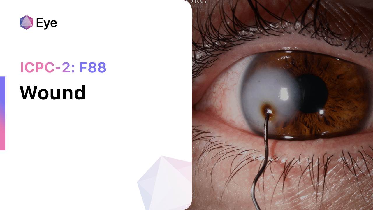
伤口 (ICPC-2: F88)
8 2 月, 2025
F86 沙眼 (ICD-10:A71)

8 2 月, 2025
F85 角膜溃疡 (ICD-10:H16.0)
8 2 月, 2025
F84 黄斑变性 (ICD-10:H35.3)
8 2 月, 2025
F83 视网膜病 (ICD-10:H35.9)
8 2 月, 2025
F82 视网膜脱离 (ICD-10:H33.0)
8 2 月, 2025
F81 先天性眼睛异常, 其他 (ICD-10:Q15.9)
8 2 月, 2025
F80 婴儿泪道阻塞 (ICD-10:Q10.5)

8 2 月, 2025
F79 眼部其他损伤 (ICD-10:S05.8)
8 2 月, 2025
F76 眼内异物 (ICD-10:T15)
7 2 月, 2025
F75 眼部挫伤/出血 (ICD-10:S05.1)
7 2 月, 2025
F74 眼部/附件肿瘤 (ICD-10:C69, D31)
7 2 月, 2025
F73 眼部感染/炎症其他 (ICD-10:H10.8)
7 2 月, 2025
F72 睑缘炎/麦粒肿/睑板腺囊肿 (ICD-10:H00, H01.0)
7 2 月, 2025
F71 过敏性结膜炎 (ICD-10:H10.1)

7 2 月, 2025
F70 传染性结膜炎 (ICD-10:H10.9)
7 2 月, 2025
F29 眼部症状/投诉其他 (ICD-10:H57.9)
7 2 月, 2025
F28 限制功能/残疾 (f) (ICD-10:Z73.6)

600 North Bridge Rd #13-01
Parkview Square, Singapore 188788
Copyright © 2023 Mirari Doctor PTE. LTD.
All rights reserved.