
介绍
视网膜病是一种影响视网膜血管的疾病,视网膜是眼后部对光敏感的组织。这是糖尿病常见的并发症,也可能由其他医疗条件或遗传因素引起。[1][2]如果不治疗,视网膜病可能导致视力丧失或失明。[3]本指南的目的是为医疗专业人员提供视网膜病诊断步骤和可能干预措施的全面概述。
代码
症状
- 视力模糊:患者可能会经历视力清晰度的逐渐或突然丧失。[4]。
- 漂浮物:可能会有黑点或细丝漂浮在视野中。[4]。
- 视野中的黑暗或空白区域:患者可能会注意到其中央或周边视野中的黑暗或空白区域。[4]。
- 夜间视物困难:患者在低光条件下可能会看到困难。[5]。
- 色觉问题:患者可能难以区分颜色。[6]。
原因
- 糖尿病:视网膜病最常由糖尿病引起,特别是如果血糖水平控制不佳。[1][7]。
- 高血压:高血压会损害视网膜的血管。[8]。
- 高胆固醇:升高的胆固醇水平会促使视网膜病的发展。[9]。
- 吸烟:吸烟增加视网膜病的风险,并可能使病情恶化。[10]。
- 遗传因素:有些人可能在遗传上易于发展视网膜病。
诊断步骤
病史
- 获取详细的病史,包括患者的糖尿病控制情况、血压水平、胆固醇水平和吸烟史的信息。
- 询问患者可能经历的任何症状,比如视力模糊或漂浮物。
- 询问有无视网膜病或其他眼部疾病的家族病史。
体格检查
- 进行全面的眼科检查,包括视力检查、眼压测量和使用检眼镜检查视网膜。
- 寻找视网膜病的迹象,比如微血管瘤、出血或棉絮斑。
- 评估患者的整体健康,包括血压测量和心血管风险因素的评估。
实验室检查
- 空腹血糖:测量患者的空腹血糖水平以评估糖尿病控制。
- 糖化血红蛋白:确定过去2-3个月的平均血糖水平。
- 血脂分析:评估患者的胆固醇和甘油三酯水平。
- 肾功能检查:检查肾功能,因为视网膜病可能与糖尿病引起的肾病相关。
- 血常规:评估贫血或其他可能影响视网膜病的血液疾病。
诊断成像
- 眼底摄影:拍摄视网膜的照片以记录视网膜病的存在和严重程度。
- 光学相干断层扫描(OCT):使用OCT获取视网膜的高分辨率横截面图像,以便详细评估视网膜厚度和液体或肿胀的存在。
- 荧光素血管造影:向患者的手臂注射荧光染料,并拍摄连续的视网膜照片以评估血流并确定渗漏或异常血管的区域。
其他检查
- 视网膜电图(ERG):测量视网膜的电反应以评估其功能。
- 视觉域测试:评估患者的周边视觉以检测任何视觉域损失的区域。
- 基因检测:考虑为有视网膜病家族史或可疑遗传原因的患者进行基因检测。
后续和患者教育
- 定期安排随访预约以监测视网膜病的进展并根据需要调整治疗。
- 教育患者了解糖尿病控制、血压管理和胆固醇控制对预防或减缓视网膜病进展的重要性。
- 鼓励患者定期进行眼科检查,并及时报告任何视力或症状的变化。
可能的干预措施
传统干预措施
药物:
视网膜病的前5种药物:
- 抗VEGF剂(如贝伐单抗、雷珠单抗):
- 费用:每次注射1800至2000美元。
- 禁忌症:对药物过敏,活动性眼部感染。
- 副作用:眼痛,眼压升高,结膜出血。
- 严重副作用:视网膜脱离,眼内炎。
- 药物相互作用:未报告。
- 警告:需要定期监测眼压和感染迹象。
- 皮质类固醇(如去炎松、地塞米松):
- 费用:每次注射500至1000美元。
- 禁忌症:活动性眼部感染,青光眼。
- 副作用:眼压升高,白内障形成。
- 严重副作用:视网膜脱离,眼内炎。
- 药物相互作用:未报告。
- 警告:需要定期监测眼压和感染迹象。
- 激光光凝术:
- 成本:每次疗程1000至2000美元。
- 禁忌症:涉及黄斑中心的黄斑水肿。
- 副作用:视力模糊,夜视能力下降。
- 严重副作用:视网膜脱离,瘢痕形成。
- 药物相互作用:未报告。
- 警告:在某些情况下可能导致永久性视力丧失。
- 玻璃体切除术:
- 费用:每次手术5000至10000美元。
- 禁忌症:严重视网膜脱离,未控制的青光眼。
- 副作用:漂浮物,白内障形成。
- 严重副作用:视网膜脱离,感染。
- 药物相互作用:未报告。
- 警告:比其他干预措施需要更长的恢复期。
- 眼内植入物(如氟烯诺酮、地塞米松):
- 费用:每次植入1500至2500美元。
- 禁忌症:活动性眼部感染,青光眼。
- 副作用:眼压升高,白内障形成。
- 严重副作用:视网膜脱离,眼内炎。
- 药物相互作用:未报告。
- 警告:需要定期监测眼压和感染迹象。
外科手术:
- 玻璃体切除术:去除玻璃体凝胶及眼中的瘢痕组织或血块以改善视力。费用:每次手术5000至10000美元。
- 视网膜脱离修复:手术重新附着视网膜到眼部后方。费用:每次手术 $5,000-$10,000。
替代干预措施
- 针灸:可能有助于改善血液流动和减少疼痛。费用:每次疗程 $60-$120。
- 螯合疗法:有争议的治疗方式,涉及给体内给予螯合剂以去除重金属。费用:每次疗程 $75-$150。
- 高压氧疗法:在加压舱内呼吸纯氧,以增加氧气输送到组织。费用:每次疗程 $200-$300。
- 草药补充剂:某些草药,如越橘和银杏叶,可能对改善视网膜健康有潜在益处。费用:具体补充剂费用因具体情况而异。
生活方式干预措施
- 血糖控制:鼓励患者通过饮食、锻炼和药物依从性来保持良好的血糖控制。
- 血压管理:建议患者通过生活方式调整与必要时的药物控制血压。
- 胆固醇控制:强调通过饮食、锻炼和需要时的药物来维持健康的胆固醇水平的重要性。
- 戒烟:鼓励患者戒烟以减少视网膜病变进展的风险。
- 定期锻炼:促进规律的身体活动,以改善整体心血管健康和增强眼部血流。
需要注意的是,提供的费用范围是近似值,其可能会根据当地情况和干预措施的可用性有所不同。
Mirari 冷等离子体替代干预
了解 Mirari 冷等离子体
- 安全且无创的治疗:Mirari 冷等离子体是一种针对多种皮肤状况的安全无创治疗选项,不需要切口,减少瘢痕、出血或组织损伤的风险。
- 有效去除异物:Mirari 冷等离子体通过降解和分解有机物质,方便从皮肤中去除异物,便于提取。
- 止痛和舒适:Mirari 冷等离子体具有局部止痛效果,在治疗过程中提供疼痛缓解,使患者更为舒适。
- 降低感染风险:Mirari 冷等离子体具有抗菌特性,有效杀菌并降低感染风险。
- 加速愈合和减少瘢痕:Mirari 冷等离子体促进伤口愈合和组织再生,缩短愈合时间并减少瘢痕的形成。
Mirari 冷等离子体处方
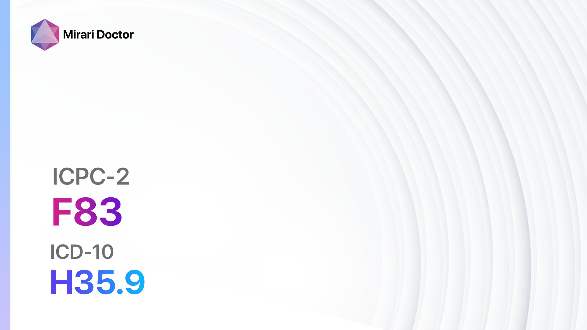
使用 Mirari 冷等离子体设备的视频说明 – F83 视网膜病变 (ICD-10:H35.9)
| 轻微 | 中度 | 重度 |
| 模式设置:1(感染) 位置:7(神经系统及耳鼻喉) 早上: 15 分钟, 晚上: 15 分钟 |
模式设置:1(感染) 位置:7(神经系统及耳鼻喉) 早上: 30 分钟, 午餐: 30 分钟, 晚上: 30 分钟 |
模式设置:1(感染) 位置:7(神经系统及耳鼻喉) 早上: 30 分钟, 午餐: 30 分钟, 晚上: 30 分钟 |
| 模式 设置: 2(伤口愈合) 位置: 7(神经系统及耳鼻喉) 早上:15 分钟, 晚上:15 分钟 |
模式 设置: 2(伤口愈合) 位置: 7(神经系统及耳鼻喉) 早上:30 分钟, 午餐:30 分钟, 晚上:30 分钟 |
模式 设置: 2(伤口愈合) 位置: 7(神经系统及耳鼻喉) 早上:30 分钟, 午餐:30 分钟, 晚上:30 分钟 |
| 模式 设置: 3(抗病毒治疗) 位置: 7(神经系统及耳鼻喉) 早上:15 分钟, 晚上:15 分钟 |
模式 设置: 3(抗病毒治疗) 位置: 7(神经系统及耳鼻喉) 早上:30 分钟, 午餐:30 分钟, 晚上:30 分钟 |
模式 设置: 3(抗病毒治疗) 位置: 7(神经系统及耳鼻喉) 早上:30 分钟, 午餐:30 分钟, 晚上:30 分钟 |
| 总计 早晨: 45 分钟 约 $7.50 美元, 晚上: 45 分钟 约 $7.50 美元 |
总计 早晨: 90 分钟 约 $15 美元, 午餐: 90 分钟 约 $15 美元, 晚上: 90 分钟 约 $15 美元, |
总计 早晨: 90 分钟 约 $15 美元, 午餐: 90 分钟 约 $15 美元, 晚上: 90 分钟 约 $15 美元, |
| 通常 治疗 持续 7-60 天 约 $105 美元 – $900 美元 | 通常 治疗 持续 6-8 周 约 $1,890 美元 – $2,520 美元 |
通常 治疗 持续 3-6 月 约 $4,050 美元 – $8,100 美元
|
 1") |
|
使用Mirari冷等离子设备有效治疗视网膜病变。
警告:Mirari冷等离子设计用于人体,不含任何人工或第三方产品。与其他产品结合使用可能导致不可预测的效果、伤害或损伤。在结合使用任何其他产品之前,请咨询专业医疗人员。
步骤1:清洁皮肤
- 首先使用温和的清洁剂或温和的肥皂和水清洁受影响的皮肤区域。用干净的毛巾轻轻拍干该区域。
步骤2:准备Mirari冷等离子设备
- 根据制造商的说明,确保Mirari冷等离子设备已完全充电或配有新电池。确保设备清洁且运行状况良好。
- 通过电源按钮或按照设备提供的具体说明打开Mirari设备。
- 某些Mirari设备可能有可调节的强度或治疗时间设置。按照制造商的说明,根据您的需要和建议的指南选择合适的设置。
步骤3:应用设备
- 将Mirari设备直接接触皮肤的受影响区域。轻轻滑动或保持设备在皮肤表面,以确保均匀覆盖需要治疗的区域。
- 缓慢以圆周运动或按照用户手册中指示的特定模式移动Mirari设备。这有助于确保全面覆盖治疗区域。
步骤4:监测和评估:
- 跟踪您的进展并评估Mirari设备在管理视网膜病变方面的效果。如果有任何疑虑或发现不良反应,请咨询您的医疗专业人员。
注意
本指南仅供信息性使用,不应替代医疗专业人员的建议。始终向您的医疗保健提供者或合格的医疗专业人员咨询以获得个人建议、诊断或治疗。请勿仅依赖此处提供的信息进行健康决策。使用此信息的风险由用户自己承担。本指南的作者及相关实体或平台不承担根据此内容产生的任何潜在的不良影响或后果的责任。
Mirari冷等离子系统免责声明
- 目的:Mirari冷等离子系统是一种 Class 2 的医疗设备,供受过培训的医疗专业人员使用。已在泰国和越南注册使用。不得在这些地点以外使用。
- 信息使用:提供的设备内容和信息仅供教育和信息之用,不能替代专业医疗建议或护理。
- 可变结果:尽管批准了该设备的特定用途,但个人结果可能不同。我们不主张或保证特定的医学结果。
- 咨询:在使用设备或基于其内容作出决定之前,必须咨询认证的Mirari远程治疗师和您的医疗保健提供者,以获得特定的协议。
- 责任:通过使用本设备,用户承认并接受所有潜在的风险。制造商或分销商不对因使用本设备而产生的任何不良反应、伤害或损害负责。
- 地理可用性:此设备已通过泰国和越南 FDA 批准用于指定用途。当前,除了泰国和越南以外,Mirari冷等离子系统不可在其他地区购买或使用。
参考文献
- Rothmann M, Nyland AH, Hammelsvang L, Petersen L, Kirketerp G, Henriksen JE. 视网膜病变的患者教育. Wiley Online Library. https://onlinelibrary.wiley.com/doi/full/10.1002/edn.179. 访问日期 2024 年 6 月 18 日。
- 视网膜病变:是什么,症状和治疗. Top Doctors. https://www.topdoctors.co.uk/medical-dictionary/retinopathy. 访问日期 2024 年 6 月 18 日。
- 视网膜病变. Drugs.com. https://www.drugs.com/health-guide/retinopathy.html. 访问日期 2024 年 6 月 18 日。
- 糖尿病性视网膜病变. 国家眼科研究所. https://www.nei.nih.gov/learn-about-eye-health/eye-conditions-and-diseases/diabetic-retinopathy. 访问日期 2024 年 6 月 18 日。
- 视网膜病变. JAMA Network. https://jamanetwork.com/journals/jama/fullarticle/208559. 访问日期 2024 年 6 月 18 日。
- 糖尿病性视网膜病变 | 临床特征. Geeky Medics. https://geekymedics.com/diabetic-retinopathy/. 访问日期 2024 年 6 月 18 日。
- 糖尿病性视网膜病变 – 症状和原因. Mayo Clinic. https://www.mayoclinic.org/diseases-conditions/diabetic-retinopathy/symptoms-causes/syc-20371611. 访问日期 2024 年 6 月 18 日。
- 识别和管理糖尿病性视网膜病变. PMC – NCBI. https://www.ncbi.nlm.nih.gov/pmc/articles/PMC3218392/. 访问日期 2024 年 6 月 18 日。
- 糖尿病视网膜病变的眼科成像:综述。PMC – NCBI。https://www.ncbi.nlm.nih.gov/pmc/articles/PMC10088017/。访问日期:2024年6月18日。
- 诊断糖尿病视网膜病变的标准工具和测试。当代验光医学。https://modernod.com/articles/2019-june/standard-tools-and-tests-fordiagnosing-diabetic-retinopathy。访问日期:2024年6月18日。
Related articles
9 2 月, 2025
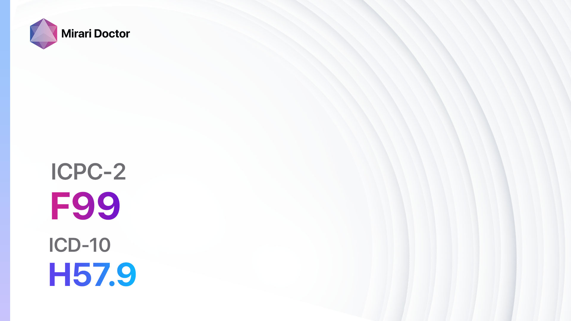
F99 眼/附件疾病,其他 (ICD-10:H57.9)
9 2 月, 2025
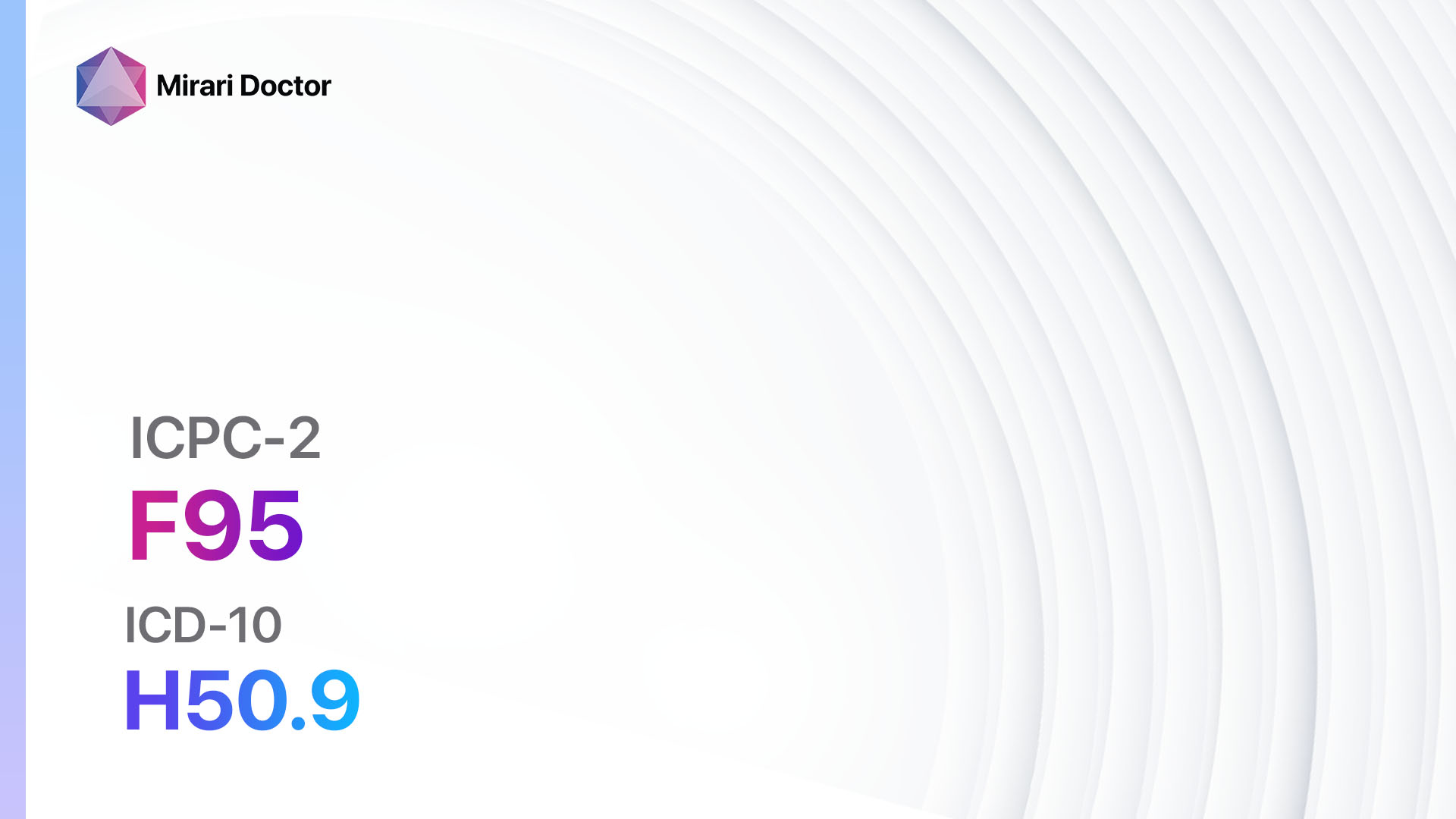
F95 斜视 (ICD-10:H50.9)

9 2 月, 2025
F94 失明 (ICD-10:H54.0)
9 2 月, 2025
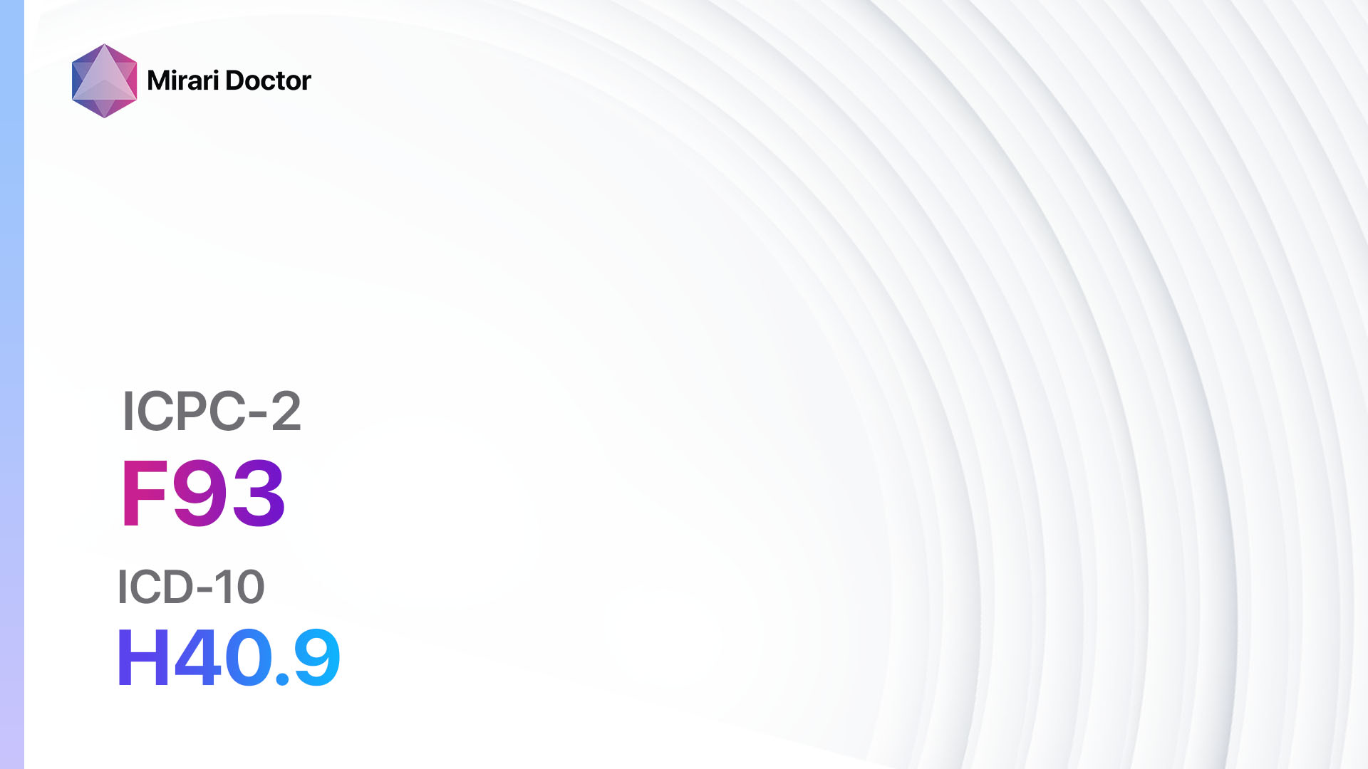
F93 青光眼 (ICD-10:H40.9)
8 2 月, 2025
F92 白内障 (ICD-10:H26.9)
8 2 月, 2025
F91 屈光不正 (ICD-10:H52.7)
8 2 月, 2025
伤口 (ICPC-2: F88)
8 2 月, 2025
F86 沙眼 (ICD-10:A71)

8 2 月, 2025
F85 角膜溃疡 (ICD-10:H16.0)
8 2 月, 2025
F84 黄斑变性 (ICD-10:H35.3)
8 2 月, 2025
F83 视网膜病 (ICD-10:H35.9)
8 2 月, 2025
F82 视网膜脱离 (ICD-10:H33.0)
8 2 月, 2025
F81 先天性眼睛异常, 其他 (ICD-10:Q15.9)
8 2 月, 2025
F80 婴儿泪道阻塞 (ICD-10:Q10.5)

8 2 月, 2025
F79 眼部其他损伤 (ICD-10:S05.8)
8 2 月, 2025
F76 眼内异物 (ICD-10:T15)
7 2 月, 2025
F75 眼部挫伤/出血 (ICD-10:S05.1)
7 2 月, 2025
F74 眼部/附件肿瘤 (ICD-10:C69, D31)
7 2 月, 2025
F73 眼部感染/炎症其他 (ICD-10:H10.8)
7 2 月, 2025
F72 睑缘炎/麦粒肿/睑板腺囊肿 (ICD-10:H00, H01.0)
7 2 月, 2025
F71 过敏性结膜炎 (ICD-10:H10.1)

7 2 月, 2025
F70 传染性结膜炎 (ICD-10:H10.9)
7 2 月, 2025
F29 眼部症状/投诉其他 (ICD-10:H57.9)
7 2 月, 2025
F28 限制功能/残疾 (f) (ICD-10:Z73.6)
600 North Bridge Rd #13-01
Parkview Square, Singapore 188788
Copyright © 2023 Mirari Doctor PTE. LTD.
All rights reserved.