Introduction
Malignant neoplasm of the nervous system refers to the presence of cancerous cells in the brain or spinal cord[1]. This condition is significant as it can lead to various neurological symptoms and can be life-threatening if not diagnosed and treated promptly[2]. The aim of this guide is to provide a comprehensive overview of the diagnostic steps, possible interventions, and patient education for malignant neoplasm of the nervous system.
Codes
- ICPC-2 Code: N74 Malignant neoplasm nervous system
- ICD-10 Code: C72.9 Malignant neoplasm of central nervous system, unspecified
Symptoms
- Headaches: Persistent or worsening headaches, especially in the morning.
- Seizures: Unexplained seizures or convulsions.
- Cognitive changes: Memory problems, confusion, or difficulty concentrating.
- Motor deficits: Weakness or numbness in the limbs.
- Vision changes: Blurred vision, double vision, or loss of peripheral vision.
- Speech difficulties: Slurred speech or difficulty finding words.
- Balance and coordination problems: Difficulty walking or maintaining balance.
- Personality changes: Mood swings, irritability, or personality changes[3].
Causes
The exact causes of malignant neoplasm of the nervous system are not fully understood. However, certain risk factors have been identified, including:
- Genetic predisposition: Some individuals may have an inherited susceptibility to developing brain tumors.
- Exposure to radiation: Previous radiation therapy to the head or neck region may increase the risk.
- Environmental factors: Exposure to certain chemicals or toxins may contribute to the development of brain tumors.
- Age: The risk of developing brain tumors increases with age, with certain types more common in specific age groups[4].
Diagnostic Steps
Medical History
- Gather information about the patient’s symptoms, including the duration, frequency, and severity.
- Identify any risk factors, such as previous radiation therapy or family history of brain tumors.
- Assess the patient’s medical history, including any previous neurological conditions or surgeries.
- Inquire about any recent changes in cognitive function, motor skills, or sensory perception.
Physical Examination
- Perform a comprehensive neurological examination to assess motor function, sensory perception, reflexes, and coordination.
- Check for any abnormalities in cranial nerve function, such as visual disturbances or facial weakness.
- Evaluate the patient’s gait and balance to identify any signs of motor deficits.
- Palpate the head and neck region to check for any lumps or abnormalities.
Laboratory Tests
- Complete blood count (CBC): To assess for any abnormalities or signs of infection.
- Liver function tests: To evaluate liver function, as liver metastases can occur in advanced cases.
- Kidney function tests: To assess renal function, as certain chemotherapy drugs may require dose adjustments.
- Tumor markers: Blood tests for specific tumor markers, such as alpha-fetoprotein (AFP) or carcinoembryonic antigen (CEA), may be useful in certain cases.
Diagnostic Imaging
- Magnetic Resonance Imaging (MRI): Provides detailed images of the brain and spinal cord to identify the location, size, and characteristics of the tumor.
- Computed Tomography (CT) scan: Can be used to visualize the brain and detect any abnormalities, such as tumors or hemorrhages.
- Positron Emission Tomography (PET) scan: Helps determine the metabolic activity of the tumor and identify any areas of spread.
- Angiography: Involves injecting a contrast dye into the blood vessels to visualize the blood supply to the tumor[5].
Other Tests
- Biopsy: In some cases, a tissue sample may be obtained through a surgical procedure or stereotactic biopsy to confirm the diagnosis and determine the tumor type.
- Lumbar puncture: Cerebrospinal fluid (CSF) analysis may be performed to check for the presence of cancer cells or other abnormalities.
- Genetic testing: In certain cases, genetic testing may be recommended to identify specific gene mutations associated with brain tumors[6].
Follow-up and Patient Education
- Schedule regular follow-up appointments to monitor the patient’s response to treatment and assess for any recurrence or progression of the tumor.
- Provide education on the signs and symptoms of tumor recurrence or treatment-related complications.
- Discuss the importance of adherence to treatment plans and any necessary lifestyle modifications.
- Offer emotional support and resources for patients and their families to cope with the diagnosis and treatment process[7].
Possible Interventions
Traditional Interventions
Medications:
Top 5 drugs for Malignant neoplasm nervous system:
- Temozolomide:
- Cost: $3,000-$5,000 per cycle.
- Contraindications: Hypersensitivity to temozolomide or its components.
- Side effects: Nausea, vomiting, fatigue, myelosuppression.
- Severe side effects: Bone marrow suppression, hepatotoxicity, secondary malignancies.
- Drug interactions: None specified.
- Warning: Regular blood tests required to monitor blood counts and liver function.
- Carmustine:
- Cost: $2,000-$4,000 per dose.
- Contraindications: Hypersensitivity to carmustine or its components.
- Side effects: Nausea, vomiting, myelosuppression, pulmonary toxicity.
- Severe side effects: Bone marrow suppression, hepatotoxicity, secondary malignancies.
- Drug interactions: None specified.
- Warning: Regular blood tests required to monitor blood counts and liver function.
- Lomustine:
- Cost: $1,500-$3,000 per dose.
- Contraindications: Hypersensitivity to lomustine or its components.
- Side effects: Nausea, vomiting, myelosuppression, hepatotoxicity.
- Severe side effects: Bone marrow suppression, hepatotoxicity, secondary malignancies.
- Drug interactions: None specified.
- Warning: Regular blood tests required to monitor blood counts and liver function.
- Bevacizumab:
- Cost: $5,000-$10,000 per dose.
- Contraindications: Hypersensitivity to bevacizumab or its components, uncontrolled hypertension.
- Side effects: Hypertension, proteinuria, bleeding, gastrointestinal perforation.
- Severe side effects: Thromboembolic events, impaired wound healing, gastrointestinal perforation.
- Drug interactions: None specified.
- Warning: Increased risk of bleeding and impaired wound healing.
- Temozolomide + Radiation Therapy:
- Cost: $10,000-$20,000 per cycle.
- Contraindications: Hypersensitivity to temozolomide or its components.
- Side effects: Nausea, vomiting, fatigue, myelosuppression.
- Severe side effects: Bone marrow suppression, hepatotoxicity, secondary malignancies.
- Drug interactions: None specified.
- Warning: Regular blood tests required to monitor blood counts and liver function.
Alternative Drugs:
- Procarbazine: An alternative chemotherapy drug that may be used in combination with other agents.
- Lomustine + Procarbazine + Vincristine: A combination chemotherapy regimen used in certain cases.
- Radiation Therapy: External beam radiation therapy may be used as the primary treatment or in combination with chemotherapy.
Surgical Procedures:
- Craniotomy: Surgical removal of the brain tumor. Cost: $50,000-$100,000.
- Stereotactic Radiosurgery: Non-invasive procedure that delivers high-dose radiation to the tumor. Cost: $30,000-$60,000.
- Shunt Placement: In cases of hydrocephalus, a shunt may be placed to drain excess cerebrospinal fluid. Cost: $20,000-$40,000.
Alternative Interventions
- Acupuncture: May help alleviate symptoms such as pain and nausea. Cost: $60-$120 per session.
- Meditation and Mindfulness: Can help reduce stress and improve overall well-being. Cost: Free to $100 for classes or workshops.
- Nutritional Therapy: A balanced diet rich in fruits, vegetables, and whole grains may support overall health. Cost: Varies depending on dietary choices.
- Herbal Supplements: Some herbal supplements, such as turmeric or green tea extract, may have potential anti-cancer properties. Cost: Varies depending on the specific supplement.
- Hyperthermia Therapy: Involves heating the tumor to destroy cancer cells. Cost: $200-$500 per session.
Lifestyle Interventions
- Regular Exercise: Physical activity can improve overall health and well-being. Cost: Free to gym membership fees.
- Stress Management: Techniques such as yoga or meditation can help reduce stress levels. Cost: Free to $100 for classes or workshops.
- Healthy Diet: A balanced diet rich in fruits, vegetables, and whole grains can support overall health. Cost: Varies depending on dietary choices.
- Smoking Cessation: Quitting smoking can improve overall health and reduce the risk of cancer recurrence. Cost: Varies depending on the method used.
- Support Groups: Joining a support group can provide emotional support and resources for coping with the diagnosis. Cost: Free to nominal fees for group membership.
It is important to note that the cost ranges provided are approximate and may vary depending on the location and availability of the interventions.
Mirari Cold Plasma Alternative Intervention
Understanding Mirari Cold Plasma
- Safe and Non-Invasive Treatment: Mirari Cold Plasma is a safe and non-invasive treatment option for various skin conditions. It does not require incisions, minimizing the risk of scarring, bleeding, or tissue damage.
- Efficient Extraction of Foreign Bodies: Mirari Cold Plasma facilitates the removal of foreign bodies from the skin by degrading and dissociating organic matter, allowing easier access and extraction.
- Pain Reduction and Comfort: Mirari Cold Plasma has a local analgesic effect, providing pain relief during the treatment, making it more comfortable for the patient.
- Reduced Risk of Infection: Mirari Cold Plasma has antimicrobial properties, effectively killing bacteria and reducing the risk of infection.
- Accelerated Healing and Minimal Scarring: Mirari Cold Plasma stimulates wound healing and tissue regeneration, reducing healing time and minimizing the formation of scars.
Mirari Cold Plasma Prescription
Video instructions for using Mirari Cold Plasma Device – N74 Malignant neoplasm nervous system (ICD-10:C72.9)
| Mild | Moderate | Severe |
| Mode setting: 1 (Infection) Location: 0 (Localized) Morning: 15 minutes, Evening: 15 minutes |
Mode setting: 1 (Infection) Location: 0 (Localized) Morning: 30 minutes, Lunch: 30 minutes, Evening: 30 minutes |
Mode setting: 1 (Infection) Location: 0 (Localized) Morning: 30 minutes, Lunch: 30 minutes, Evening: 30 minutes |
| Mode setting: 2 (Wound Healing) Location: 0 (Localized) Morning: 15 minutes, Evening: 15 minutes |
Mode setting: 2 (Wound Healing) Location: 0 (Localized) Morning: 30 minutes, Lunch: 30 minutes, Evening: 30 minutes |
Mode setting: 2 (Wound Healing) Location: 0 (Localized) Morning: 30 minutes, Lunch: 30 minutes, Evening: 30 minutes |
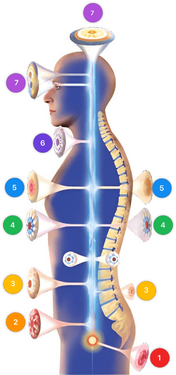
| Mode setting: 7 (Immunotherapy) Location: 1 (Sacrum) Morning: 15 minutes, Evening: 15 minutes |
Mode setting: 7 (Immunotherapy) Location: 1 (Sacrum) Morning: 30 minutes, Lunch: 30 minutes, Evening: 30 minutes |
Mode setting: 7 (Immunotherapy) Location: 1 (Sacrum) Morning: 30 minutes, Lunch: 30 minutes, Evening: 30 minutes |
| Total Morning: 60 minutes approx. $10 USD, Evening: 60 minutes approx. $10 USD |
Total Morning: 120 minutes approx. $20 USD, Lunch: 120 minutes approx. $20 USD, Evening: 120 minutes approx. $20 USD, |
Total Morning: 120 minutes approx. $20 USD, Lunch: 120 minutes approx. $20 USD, Evening: 120 minutes approx. $20 USD, |
| Usual treatment for 7-60 days approx. $140 USD – $1200 USD | Usual treatment for 6-8 weeks approx. $2,520 USD – $3,360 USD |
Usual treatment for 3-6 months approx. $5,400 USD – $10,800 USD
|
 |
|
Use the Mirari Cold Plasma device to treat Malignant neoplasm nervous system effectively.
WARNING: MIRARI COLD PLASMA IS DESIGNED FOR THE HUMAN BODY WITHOUT ANY ARTIFICIAL OR THIRD PARTY PRODUCTS. USE OF OTHER PRODUCTS IN COMBINATION WITH MIRARI COLD PLASMA MAY CAUSE UNPREDICTABLE EFFECTS, HARM OR INJURY. PLEASE CONSULT A MEDICAL PROFESSIONAL BEFORE COMBINING ANY OTHER PRODUCTS WITH USE OF MIRARI.[8][9][10]
Step 1: Cleanse the Skin
- Start by cleaning the affected area of the skin with a gentle cleanser or mild soap and water. Gently pat the area dry with a clean towel.
Step 2: Prepare the Mirari Cold Plasma device
- Ensure that the Mirari Cold Plasma device is fully charged or has fresh batteries as per the manufacturer’s instructions. Make sure the device is clean and in good working condition.
- Switch on the Mirari device using the power button or by following the specific instructions provided with the device.
- Some Mirari devices may have adjustable settings for intensity or treatment duration. Follow the manufacturer’s instructions to select the appropriate settings based on your needs and the recommended guidelines.
Step 3: Apply the Device
- Place the Mirari device in direct contact with the affected area of the skin. Gently glide or hold the device over the skin surface, ensuring even coverage of the area experiencing.
- Slowly move the Mirari device in a circular motion or follow a specific pattern as indicated in the user manual. This helps ensure thorough treatment coverage.
Step 4: Monitor and Assess:
- Keep track of your progress and evaluate the effectiveness of the Mirari device in managing your Malignant neoplasm nervous system. If you have any concerns or notice any adverse reactions, consult with your health care professional.
Note
This guide is for informational purposes only and should not replace the advice of a medical professional. Always consult with your healthcare provider or a qualified medical professional for personal advice, diagnosis, or treatment. Do not solely rely on the information presented here for decisions about your health. Use of this information is at your own risk. The authors of this guide, nor any associated entities or platforms, are not responsible for any potential adverse effects or outcomes based on the content.
Mirari Cold Plasma System Disclaimer
- Purpose: The Mirari Cold Plasma System is a Class 2 medical device designed for use by trained healthcare professionals. It is registered for use in Thailand and Vietnam. It is not intended for use outside of these locations.
- Informational Use: The content and information provided with the device are for educational and informational purposes only. They are not a substitute for professional medical advice or care.
- Variable Outcomes: While the device is approved for specific uses, individual outcomes can differ. We do not assert or guarantee specific medical outcomes.
- Consultation: Prior to utilizing the device or making decisions based on its content, it is essential to consult with a Certified Mirari Tele-Therapist and your medical healthcare provider regarding specific protocols.
- Liability: By using this device, users are acknowledging and accepting all potential risks. Neither the manufacturer nor the distributor will be held accountable for any adverse reactions, injuries, or damages stemming from its use.
- Geographical Availability: This device has received approval for designated purposes by the Thai and Vietnam FDA. As of now, outside of Thailand and Vietnam, the Mirari Cold Plasma System is not available for purchase or use.
References
- Ostrom QT, Patil N, Cioffi G, Waite K, Kruchko C, Barnholtz-Sloan JS. CBTRUS Statistical Report: Primary Brain and Other Central Nervous System Tumors Diagnosed in the United States in 2013-2017. Neuro Oncol. 2020;22(12 Suppl 2):iv1-iv96. doi:10.1093/neuonc/noaa200
- Lapointe S, Perry A, Butowski NA. Primary brain tumours in adults. Lancet. 2018;392(10145):432-446. doi:10.1016/S0140-6736(18)30990-5
- Weller M, van den Bent M, Preusser M, et al. EANO guidelines on the diagnosis and treatment of diffuse gliomas of adulthood. Nat Rev Clin Oncol. 2021;18(3):170-186. doi:10.1038/s41571-020-00447-z
- Ostrom QT, Gittleman H, Truitt G, Boscia A, Kruchko C, Barnholtz-Sloan JS. CBTRUS Statistical Report: Primary Brain and Other Central Nervous System Tumors Diagnosed in the United States in 2011-2015. Neuro Oncol. 2018;20(suppl_4):iv1-iv86. doi:10.1093/neuonc/noy131
- Huang RY, Neagu MR, Reardon DA, Wen PY. Pitfalls in the neuroimaging of glioblastoma in the era of antiangiogenic and immuno/targeted therapy – detecting illusive disease, defining response. Front Neurol. 2015;6:33. doi:10.3389/fneur.2015.00033
- Eckel-Passow JE, Lachance DH, Molinaro AM, et al. Glioma Groups Based on 1p/19q, IDH, and TERT Promoter Mutations in Tumors. N Engl J Med. 2015;372(26):2499-2508. doi:10.1056/NEJMoa1407279
- Langbecker D, Yates P. Primary brain tumor patients’ supportive care needs and multidisciplinary rehabilitation, community and psychosocial support services: awareness, referral and utilization. J Neurooncol. 2016;127(1):91-102. doi:10.1007/s11060-015-2013-9
- Skvara, Hans ; Teban, Ligia ; Fiebiger, Manfred ; Binder, Michael ; Kittler, Harald (2005). Limitations of Dermoscopy in the Recognition of Melanoma. DOI: 10.1001/archderm.141.2.155.
- Fitzpatrick, Thomas B (1988). The Validity and Practicality of Sun-Reactive Skin Types I Through VI. DOI: 10.1001/archderm.1988.01670060015008.
- Watts, Caroline G ; Cust, Anne E ; Menzies, Scott W ; Coates, Elliot ; Mann, Graham J ; Morton, Rachael L (2015). Specialized Surveillance for Individuals at High Risk for Melanoma: A Cost Analysis of a High-Risk Clinic. DOI: 10.1001/jamadermatol.2014.1952.
Related articles
Made in USA


